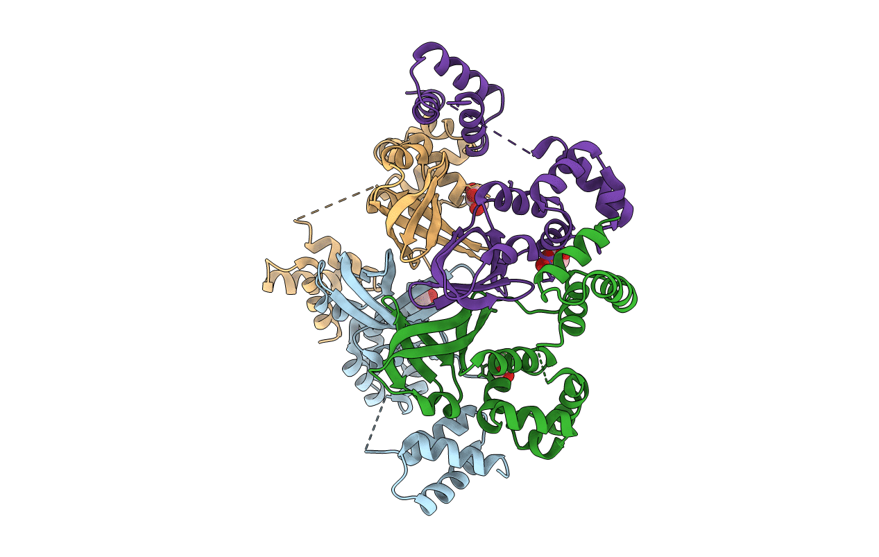
2ZTC image
Entry Detail
PDB ID:
2ZTC
Title:
MtRuvA Form II
Biological Source:
Host Organism:
PDB Version:
Deposition Date:
2008-10-01
Release Date:
2009-05-05
Method Details:
Experimental Method:
Resolution:
2.80 Å
R-Value Free:
0.28
R-Value Work:
0.22
R-Value Observed:
0.22
Space Group:
C 1 2 1
Macromolecular Entities
Polymer Type:polypeptide(L)
Description:Holliday junction ATP-dependent DNA helicase ruvA
Chain IDs:A, B, C, D
Chain Length:212
Number of Molecules:4
Biological Source:Mycobacterium tuberculosis
Ligand Molecules
Primary Citation
Crystallographic and modelling studies on Mycobacterium tuberculosis RuvA Additional role of RuvB-binding domain and inter species variability
Biochim.Biophys.Acta 1794 1001 1009 (2009)
PMID: 19374958 DOI: 10.1016/j.bbapap.2009.04.003

Abstact

RuvA, along with RuvB, is involved in branch migration of heteroduplex DNA in homologous recombination. The structures of three new crystal forms of RuvA from Mycobacterium tuberculosis (MtRuvA) have been determined. The RuvB-binding domain is cleaved off in one of them. Detailed models of the complexes of octameric RuvA from different species with the Holliday junction have also been constructed. A thorough examination of the structures presented here and those reported earlier brings to light the hitherto unappreciated role of the RuvB-binding domain in determining inter-domain orientation and oligomerization. These structures also permit an exploration of the interspecies variability of structural features such as oligomerization and the conformation of the loop that carries the acidic pin, in terms of amino acid substitutions. These models emphasize the additional role of the RuvB-binding domain in Holliday junction binding. This role along with its role in oligomerization could have important biological implications.

Legend

Protein

Chemical

Disease

Primary Citation of related structures