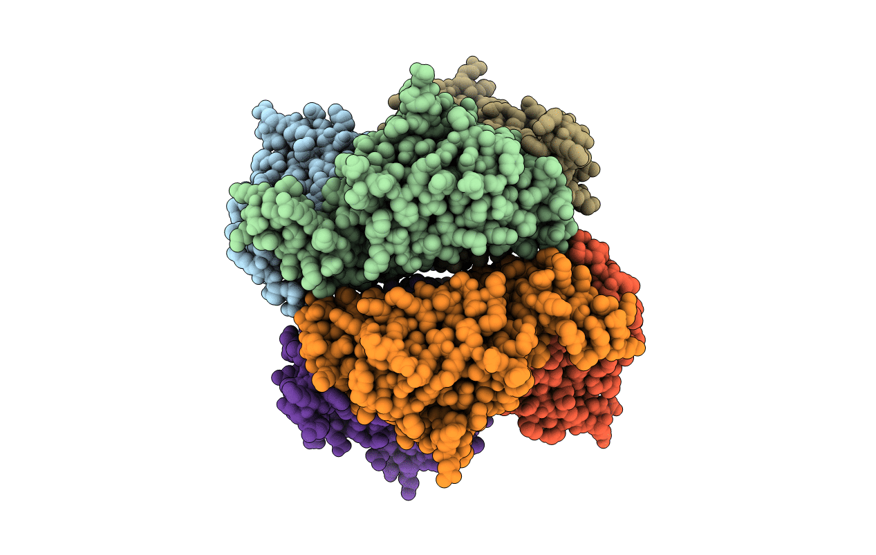
2ZQQ image
Deposition Date 2008-08-16
Release Date 2009-06-30
Last Version Date 2023-11-01
Entry Detail
PDB ID:
2ZQQ
Keywords:
Title:
Crystal structure of human AUH (3-methylglutaconyl-coa hydratase) mixed with (AUUU)24A RNA
Biological Source:
Source Organism:
Homo sapiens (Taxon ID: 9606)
Host Organism:
Method Details:
Experimental Method:
Resolution:
2.20 Å
R-Value Free:
0.24
R-Value Work:
0.20
R-Value Observed:
0.20
Space Group:
P 1 21 1
Macromolecular Entities
Structures with similar UniProt ID
Protein Blast
Polymer Type:polypeptide(L)
Molecule:Methylglutaconyl-CoA hydratase
Gene (Uniprot):AUH
Chain IDs:A, B, C, D, E, F
Chain Length:272
Number of Molecules:6
Biological Source:Homo sapiens
Primary Citation
AU-rich RNA-binding induces changes in the quaternary structure of AUH
Proteins 75 360 372 (2009)
PMID: 18831052 DOI: 10.1002/prot.22246

Abstact

The human AU RNA binding protein/enoyl-Coenzyme A hydratase (AUH) is a 3-hydroxy-3-methylglutaconyl-CoA dehydratase in the leucine degradation pathway. It also possesses an RNA-binding activity to AUUU repeats, which involves no known conserved RNA-binding domains and is seemingly unrelated to the enzymatic activity. In this study, we performed mass spectrometric analyses to elucidate the oligomeric states of AUH in the presence and absence of RNA. With a short RNA (AUUU) or without RNA, AUH mainly exists as a trimer in solution. On the other hand, the AUH trimer dimerizes upon binding to one molecule of a long RNA containing 24 repeats of the AUUU motif, (AUUU)(24)A. AUH was crystallized with the long RNA. Although the RNA was disordered in the crystalline lattice, the AUH structure was determined as an asymmetric dimer of trimers with a kink in the alignment of the trimer axes, resulting in the formation of two clefts with significantly different sizes.

Legend

Protein

Chemical

Disease

Primary Citation of related structures