Deposition Date
2008-07-31
Release Date
2008-10-28
Last Version Date
2023-11-01
Entry Detail
Biological Source:
Source Organism(s):
Bacteroides thetaiotaomicron (Taxon ID: 818)
Expression System(s):
Method Details:
Experimental Method:
Resolution:
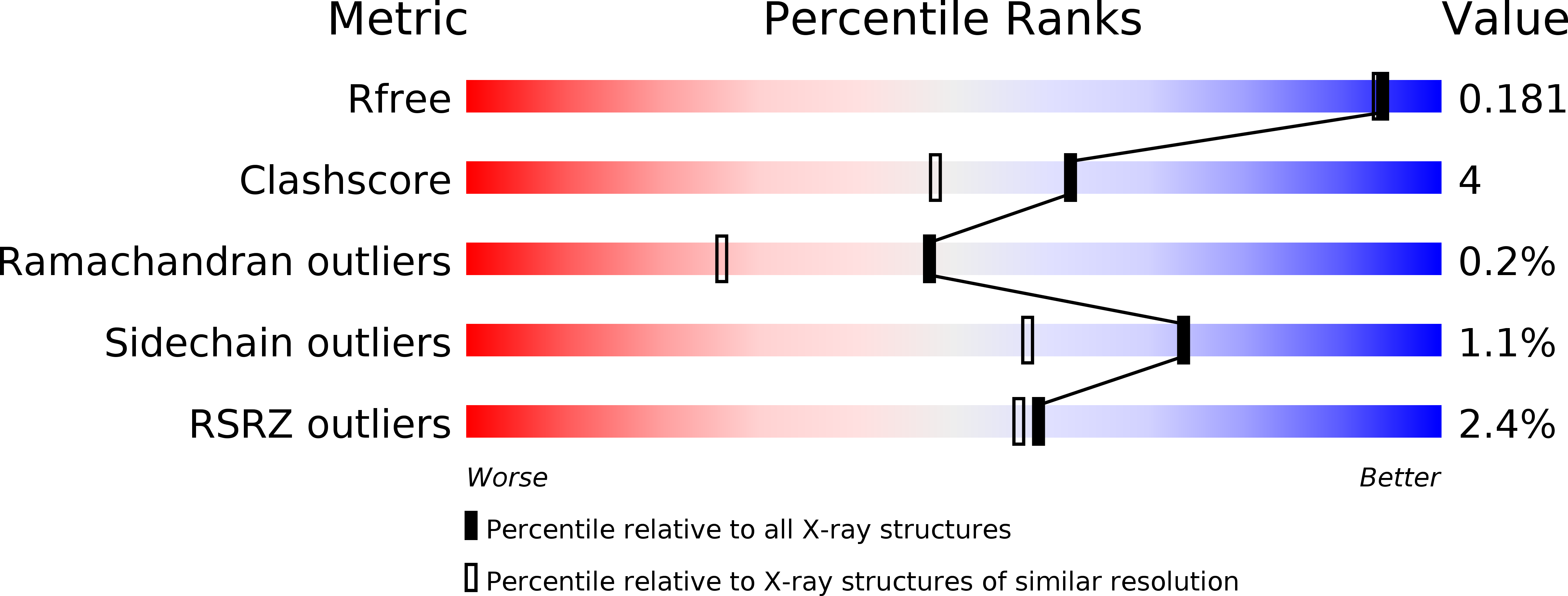
1.60 Å
R-Value Free:
0.18
R-Value Work:
0.17
Space Group:
P 1 21 1