2ZPS image
Deposition Date 2008-07-28
Release Date 2009-07-28
Last Version Date 2024-10-30
Entry Detail
PDB ID:
2ZPS
Keywords:
Title:
Crystal structure of anionic trypsin isoform 3 from chum salmon
Biological Source:
Source Organism:
Method Details:
Experimental Method:
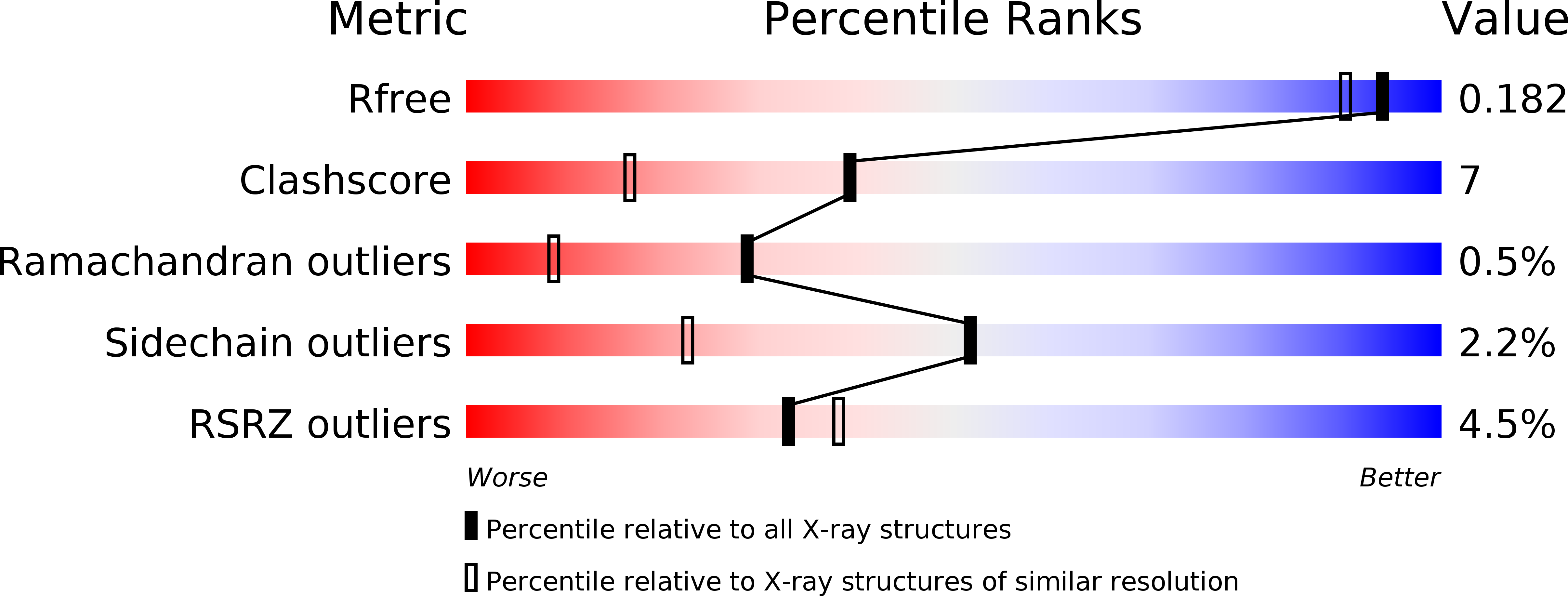
Resolution:
1.55 Å
R-Value Free:
0.19
R-Value Work:
0.18
Space Group:
P 21 21 21
Macromolecular Entities
Structures with similar UniProt ID
Protein Blast
Polymer Type:polypeptide(L)
Molecule:Anionic trypsin
Chain IDs:A
Chain Length:222
Number of Molecules:1
Biological Source:Oncorhynchus keta
Primary Citation
A structural comparison of three isoforms of anionic trypsin from chum salmon (Oncorhynchus keta).
Acta Crystallogr.,Sect.D 65 717 723 (2009)
PMID: 19564692 DOI: 10.1107/S0907444909012165

Abstact

Three anionic salmon trypsin isoforms (CST-1, CST-2 and CST-3) were isolated from the pyloric caeca of chum salmon (Oncorhynchus keta). The order of catalytic efficiency (K(m)/k(cat)) of the isoforms during BAPA hydrolysis was CST-2 > CST-1 > CST-3. In order to find a structural rationalization for the observed difference in catalytic efficiency, the X-ray crystallographic structures of the three isoforms were compared in detail. Some structural differences were observed in the C-terminal alpha-helix, interdomain loop and active-site region. From the results of the detailed comparison, it appears that the structural flexibility of the C-terminal alpha-helix, which interacts with the N-terminal domain, and the substrate-binding pocket in CST-3 are lower than those in CST-1 and CST-2. In addition, the conformation of the catalytic triad (His57, Asp102 and Ser195) differs among the three isoforms. The imidazole N atom of His57 in CST-1 and CST-2 forms a hydrogen bond to the hydroxyl O atom of Ser195, but the distance between the imidazole N atom of His57 and the hydroxyl O atom of Ser195 in CST-3 is too great (3.8 A) for the formation of a hydrogen bond. Thus, the nucleophilicity of the hydroxyl group of Ser195 in CST-3 is weaker than that in CST-1 or CST-2. Furthermore, the electrostatic potential of the substrate-binding pocket in CST-2 is markedly lower than those in CST-1 and CST-3 owing to the negative charges of Asp150, Asp153 and Glu221B that arise from the long-range effect. These results may explain the higher catalytic efficiency of CST-2 compared with CST-1 and CST-3.

Legend

Protein

Chemical

Disease

Primary Citation of related structures