2ZOF image
Deposition Date 2008-05-14
Release Date 2008-06-10
Last Version Date 2024-03-13
Entry Detail
PDB ID:
2ZOF
Keywords:
Title:
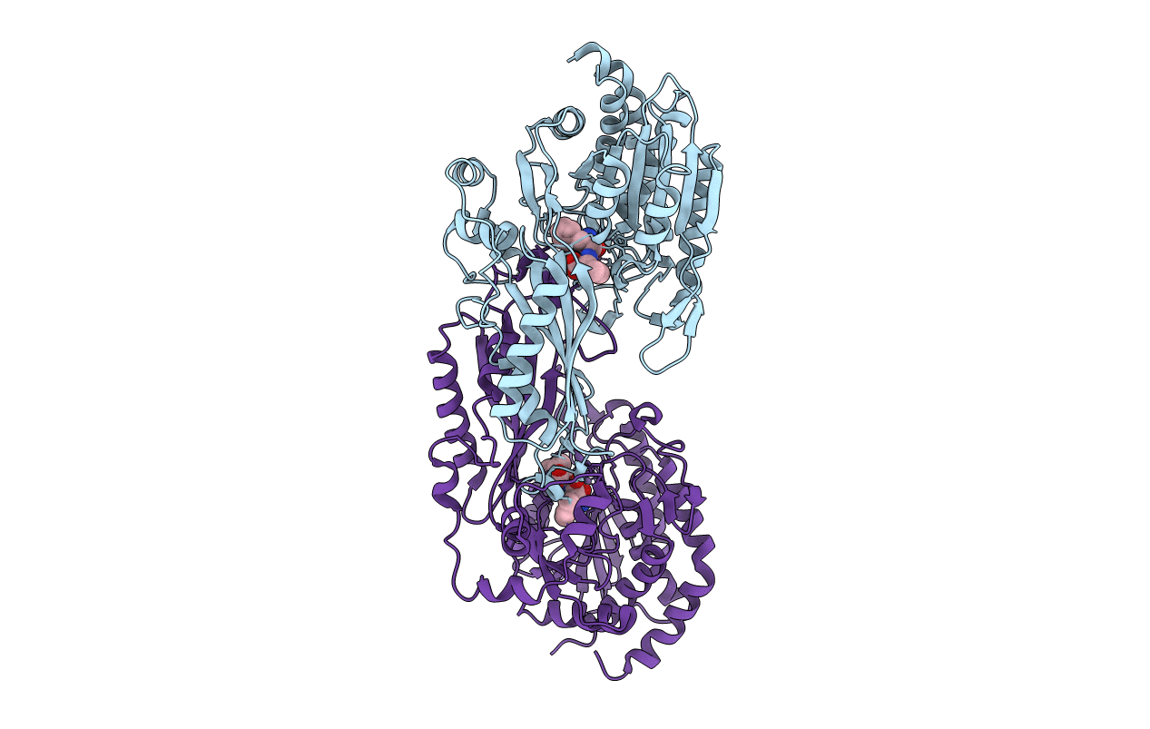
Crystal structure of mouse carnosinase CN2 complexed with MN and bestatin
Biological Source:
Source Organism:
Mus musculus (Taxon ID: 10090)
Host Organism:
Method Details:
Experimental Method:
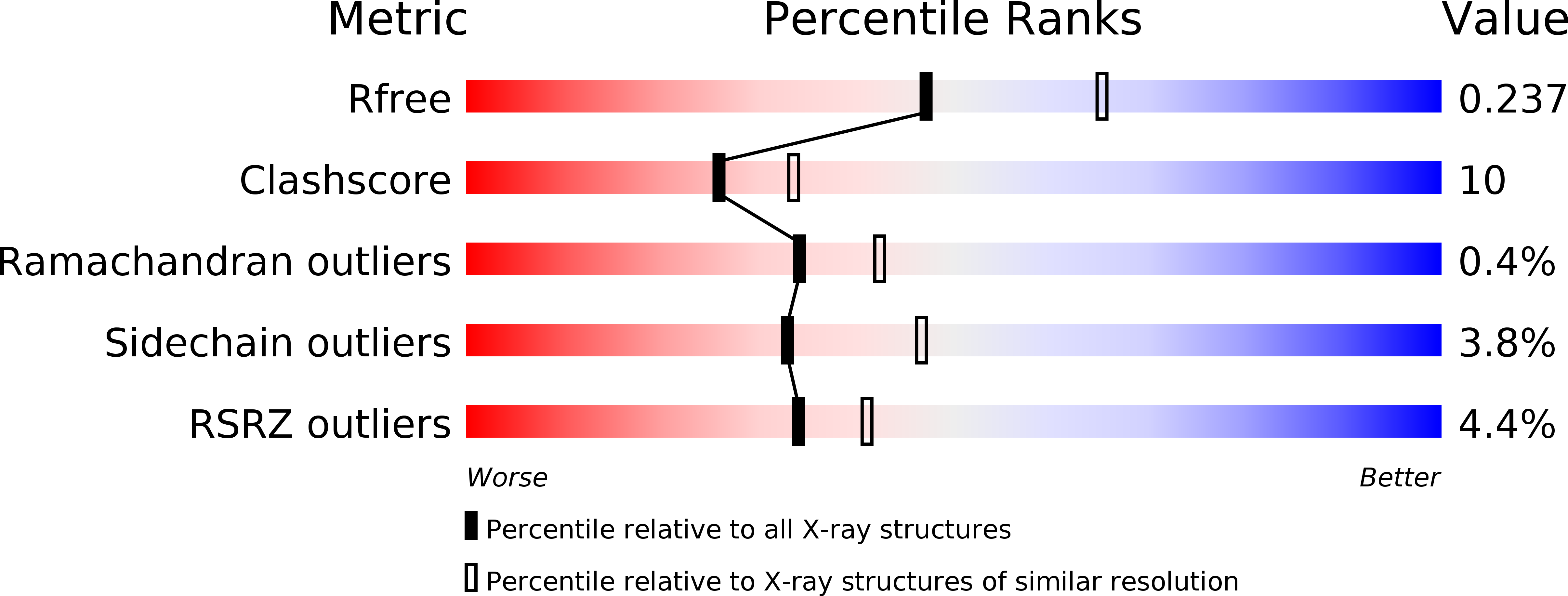
Resolution:
2.30 Å
R-Value Free:
0.23
R-Value Work:
0.19
R-Value Observed:
0.19
Space Group:
P 1 21 1
Macromolecular Entities
Structures with similar UniProt ID
Protein Blast
Polymer Type:polypeptide(L)
Molecule:Cytosolic non-specific dipeptidase
Gene (Uniprot):Cndp2
Chain IDs:A, B
Chain Length:479
Number of Molecules:2
Biological Source:Mus musculus
Primary Citation
Structural basis for substrate recognition and hydrolysis by mouse carnosinase CN2.
J.Biol.Chem. 283 27289 27299 (2008)
PMID: 18550540 DOI: 10.1074/jbc.M801657200

Abstact

L-carnosine is a bioactive dipeptide (beta-alanyl-L-histidine) present in mammalian tissues, including the central nervous system, and has potential neuroprotective and neurotransmitter functions. In mammals, two types of L-carnosine-hydrolyzing enzymes (CN1 and CN2) have been cloned thus far, and they have been classified as metallopeptidases of the M20 family. The enzymatic activity of CN2 requires Mn(2+), and CN2 is inhibited by a nonhydrolyzable substrate analog, bestatin. Here, we present the crystal structures of mouse CN2 complexed with bestatin together with Zn(2+) at a resolution of 1.7 A and that with Mn(2+) at 2.3 A CN2 is a homodimer in a noncrystallographic asymmetric unit, and the Mn(2+) and Zn(2+) complexes closely resemble each other in the overall structure. Each subunit is composed of two domains: domain A, which is complexed with bestatin and two metal ions, and domain B, which provides the major interface for dimer formation. The bestatin molecule bound to domain A interacts with several residues of domain B of the other subunit, and these interactions are likely to be essential for enzyme activity. Since the bestatin molecule is not accessible to the bulk water, substrate binding would require conformational flexibility between domains A and B. The active site structure and substrate-binding model provide a structural basis for the enzymatic activity and substrate specificity of CN2 and related enzymes.

Legend

Protein

Chemical

Disease

Primary Citation of related structures