Deposition Date
2008-04-28
Release Date
2008-09-02
Last Version Date
2024-03-13
Entry Detail
PDB ID:
2ZNL
Keywords:
Title:
Crystal structure of PA-PB1 complex form influenza virus RNA polymerase
Biological Source:
Source Organism(s):
Influenza A virus (Taxon ID: 211044)
Expression System(s):
Method Details:
Experimental Method:
Resolution:
2.30 Å
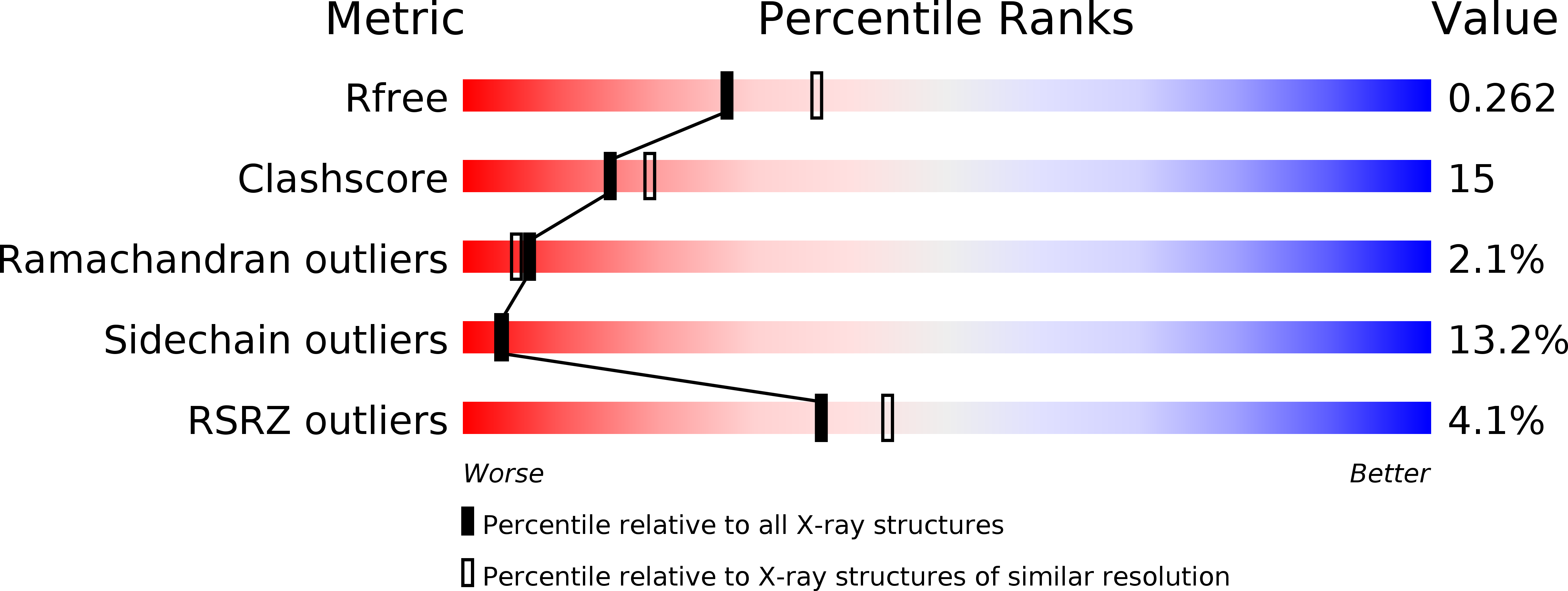
R-Value Free:
0.26
R-Value Work:
0.20
R-Value Observed:
0.20
Space Group:
P 32 2 1