2ZMX image
Entry Detail
PDB ID:
2ZMX
Title:
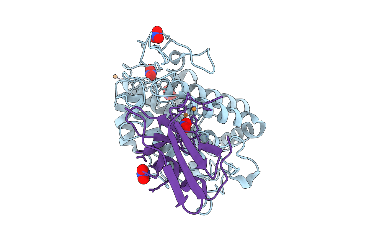
Crystal structure of the met1-form of the copper-bound tyrosinase in complex with a caddie protein from Streptomyces castaneoglobisporus obtained by soaking in cupric sulfate solution for 36 hours
Biological Source:
Host Organism:
PDB Version:
Deposition Date:
2008-04-21
Release Date:
2009-04-07
Method Details:
Experimental Method:
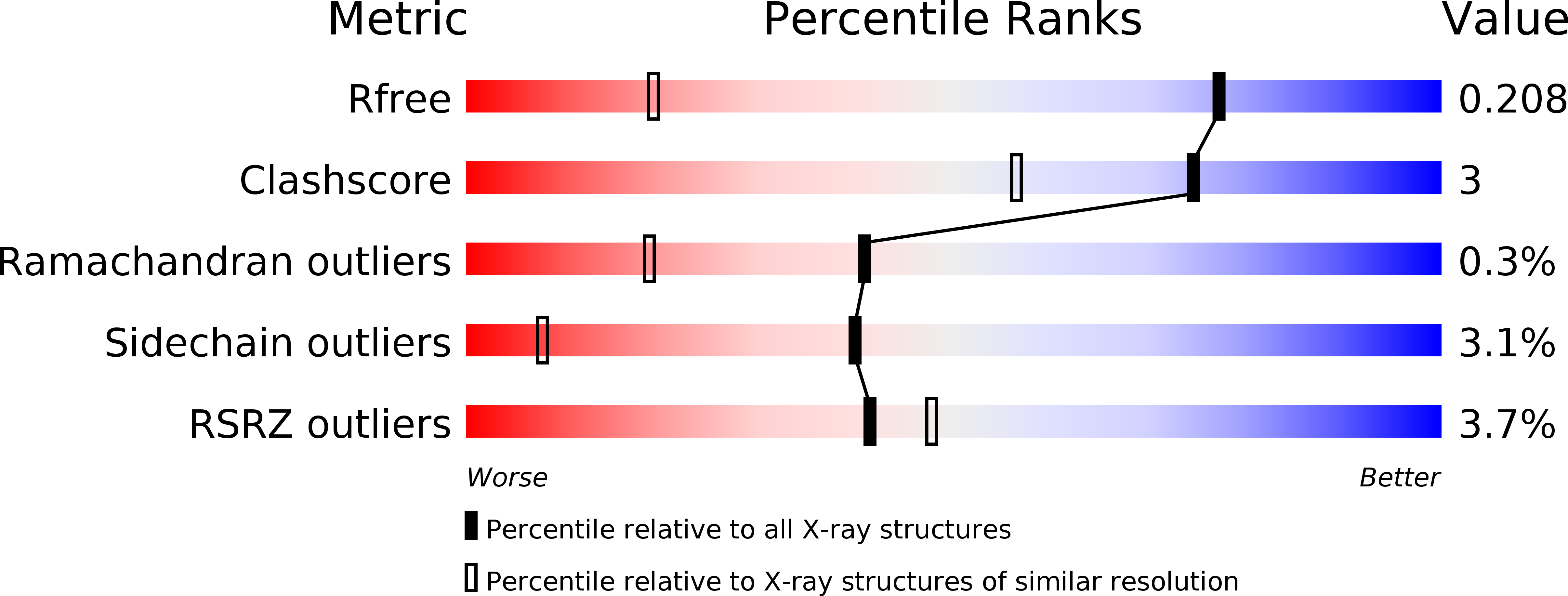
Resolution:
1.33 Å
R-Value Free:
0.21
R-Value Work:
0.17
R-Value Observed:
0.17
Space Group:
P 21 21 2
Macromolecular Entities
Polymer Type:polypeptide(L)
Description:Tyrosinase
Chain IDs:A
Chain Length:281
Number of Molecules:1
Biological Source:Streptomyces castaneoglobisporus
Polymer Type:polypeptide(L)
Description:CADDIE
Chain IDs:B
Chain Length:134
Number of Molecules:1
Biological Source:STREPTOMYCES CASTANEOGLOBISPORUS
Primary Citation
Crystallographic Evidence That the Dinuclear Copper Center of Tyrosinase Is Flexible during Catalysis
J.Biol.Chem. 281 8981 8990 (2006)
PMID: 16436386 DOI: 10.1074/jbc.M509785200

Abstact

At high resolution, we determined the crystal structures of copper-bound and metal-free tyrosinase in a complex with ORF378 designated as a "caddie" protein because it assists with transportation of two CuII ions into the tyrosinase catalytic center. These structures suggest that the caddie protein covers the hydrophobic molecular surface of tyrosinase and interferes with the binding of a substrate tyrosine to the catalytic site of tyrosinase. The caddie protein, which consists of one six-strandedbeta-sheet and one alpha-helix, has no similarity with all proteins deposited into the Protein Data Bank. Although tyrosinase and catechol oxidase are classified into the type 3 copper protein family, the latter enzyme lacks monooxygenase activity. The difference in catalytic activity is based on the structural observations that a large vacant space is present just above the active center of tyrosinase and that one of the six His ligands for the two copper ions is highly flexible. These structural characteristics of tyrosinase suggest that, in the reaction that catalyzes the ortho-hydroxylation of monophenol, one of the two Cu(II) ions is coordinated by the peroxide-originated oxygen bound to the substrate. Our crystallographic study shows evidence that the tyrosinase active center formed by dinuclear coppers is flexible during catalysis.

Legend

Protein

Chemical

Disease

Primary Citation of related structures