Deposition Date
2008-04-21
Release Date
2008-10-21
Last Version Date
2024-10-16
Entry Detail
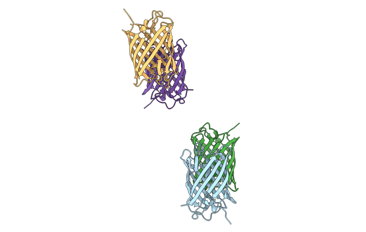
PDB ID:
2ZMW
Keywords:
Title:
Crystal Structure of Monomeric Kusabira-Orange (MKO), Orange-Emitting GFP-like Protein, at pH 6.0
Biological Source:
Source Organism(s):
Fungia concinna (Taxon ID: 496660)
Expression System(s):
Method Details:
Experimental Method:
Resolution:
2.00 Å
R-Value Free:
0.23
R-Value Work:
0.19
R-Value Observed:
0.19
Space Group:
P 1 21 1