2ZMF image
Deposition Date 2008-04-17
Release Date 2008-04-29
Last Version Date 2024-10-16
Entry Detail
PDB ID:
2ZMF
Keywords:
Title:
Crystal structure of the C-terminal GAF domain of human phosphodiesterase 10A
Biological Source:
Source Organism:
Homo sapiens (Taxon ID: 9606)
Method Details:
Experimental Method:
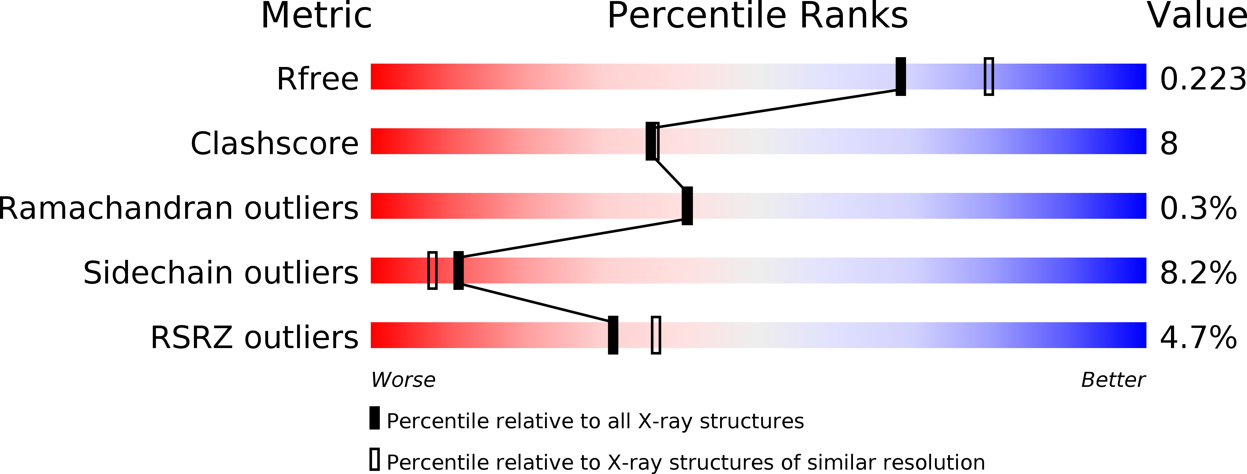
Resolution:
2.10 Å
R-Value Free:
0.22
R-Value Work:
0.19
R-Value Observed:
0.19
Space Group:
P 31 2 1
Macromolecular Entities
Polymer Type:polypeptide(L)
Molecule:cAMP and cAMP-inhibited cGMP 3',5'-cyclic phosphodiesterase 10A
Gene (Uniprot):PDE10A
Chain IDs:A, B
Chain Length:189
Number of Molecules:2
Biological Source:Homo sapiens
Modified Residue
Compound ID Chain ID Parent Comp ID Details 2D Image
MSE A MET SELENOMETHIONINE
Ligand Molecules
Primary Citation
Crystal Structure of the GAF-B Domain from Human Phosphodiesterase 10A Complexed with Its Ligand, cAMP
J.Biol.Chem. 283 19657 19664 (2008)
PMID: 18477562 DOI: 10.1074/jbc.M800595200

Abstact

Cyclic nucleotide phosphodiesterases (PDEs) catalyze the degradation of the cyclic nucleotides cAMP and cGMP, which are important second messengers. Five of the 11 mammalian PDE families have tandem GAF domains at their N termini. PDE10A may be the only mammalian PDE for which cAMP is the GAF domain ligand, and it may be allosterically stimulated by cAMP. PDE10A is highly expressed in striatal medium spiny neurons. Here we report the crystal structure of the C-terminal GAF domain (GAF-B) of human PDE10A complexed with cAMP at 2.1-angstroms resolution. The conformation of the PDE10A GAF-B domain monomer closely resembles those of the GAF domains of PDE2A and the cyanobacterium Anabaena cyaB2 adenylyl cyclase, except for the helical bundle consisting of alpha1, alpha2, and alpha5. The PDE10A GAF-B domain forms a dimer in the crystal and in solution. The dimerization is mainly mediated by hydrophobic interactions between the helical bundles in a parallel arrangement, with a large buried surface area. In the PDE10A GAF-B domain, cAMP tightly binds to a cNMP-binding pocket. The residues in the alpha3 and alpha4 helices, the beta6 strand, the loop between 3(10) and alpha4, and the loop between alpha4 and beta5 are involved in the recognition of the phosphate and ribose moieties. This recognition mode is similar to those of the GAF domains of PDE2A and cyaB2. In contrast, the adenine base is specifically recognized by the PDE10A GAF-B domain in a unique manner, through residues in the beta1 and beta2 strands.

Legend

Protein

Chemical

Disease

Primary Citation of related structures