2ZKZ image
Deposition Date 2008-04-01
Release Date 2009-04-07
Last Version Date 2024-11-13
Entry Detail
PDB ID:
2ZKZ
Keywords:
Title:
Crystal structure of the transcriptional repressor PagR of Bacillus anthracis
Biological Source:
Source Organism:
Host Organism:
Method Details:
Experimental Method:
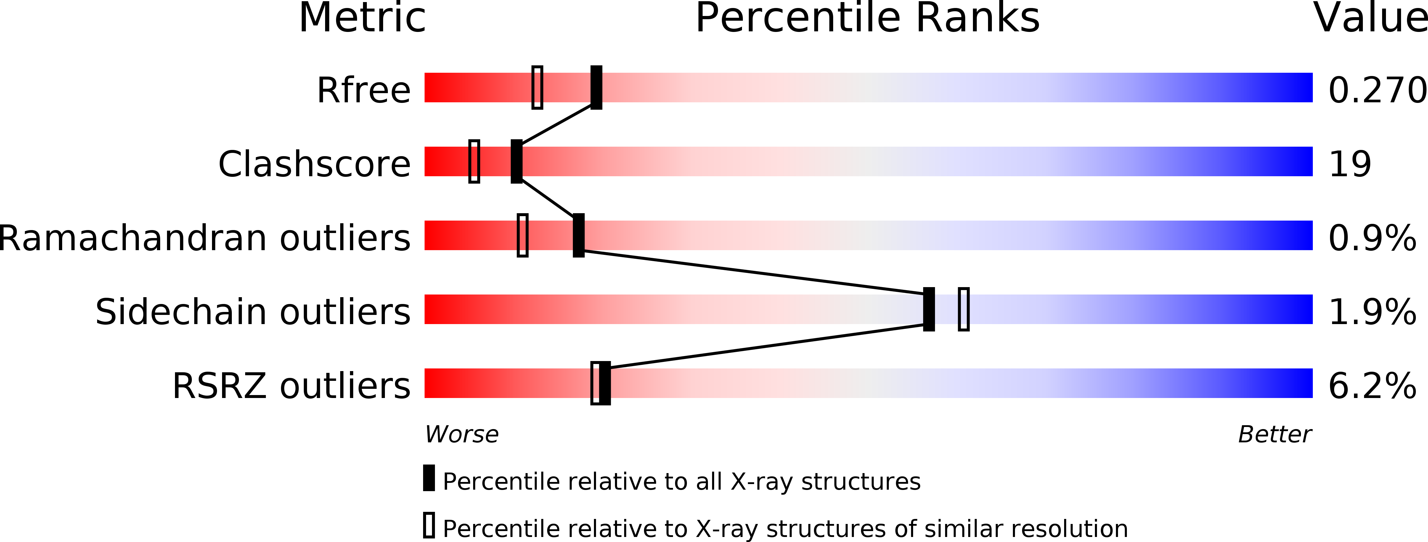
Resolution:
2.00 Å
R-Value Free:
0.26
R-Value Work:
0.24
Space Group:
P 41 21 2
Macromolecular Entities
Structures with similar UniProt ID
Protein Blast
Polymer Type:polypeptide(L)
Molecule:Transcriptional repressor pagR
Gene (Uniprot):pagR
Chain IDs:A, B, C, D
Chain Length:99
Number of Molecules:4
Biological Source:Bacillus anthracis
Modified Residue
Compound ID Chain ID Parent Comp ID Details 2D Image
MSE A MET SELENOMETHIONINE
Primary Citation
Crystal structure of the transcriptional repressor PagR of Bacillus anthracis
Microbiology 156 385 391 (2010)
PMID: 19926656 DOI: 10.1099/mic.0.033548-0

Abstact

PagR is a transcriptional repressor in Bacillus anthracis that controls the chromosomal S-layer genes eag and sap, and downregulates the protective antigen pagA gene by direct binding to their promoter regions. The PagR protein sequence is similar to those of members of the ArsR repressor family involved in the repression of arsenate-resistance genes in numerous bacteria. The crystal structure of PagR was solved using multi-wavelength anomalous diffraction (MAD) techniques and was refined with 1.8 A resolution diffraction data. The PagR molecules form dimers, as observed in all SmtB/ArsR repressor family proteins. In the crystal lattice four PagR dimers pack together to form an inactive octamer. Model-building studies suggest that the dimer binds to a DNA duplex with a bend of around 4 degrees.

Legend

Protein

Chemical

Disease

Primary Citation of related structures