2ZKD image
Deposition Date 2008-03-19
Release Date 2008-09-09
Last Version Date 2023-11-01
Entry Detail
PDB ID:
2ZKD
Keywords:
Title:
Crystal structure of the SRA domain of mouse Np95 in complex with hemi-methylated CpG DNA
Biological Source:
Source Organism:
Mus musculus (Taxon ID: 10090)
Host Organism:
Method Details:
Experimental Method:
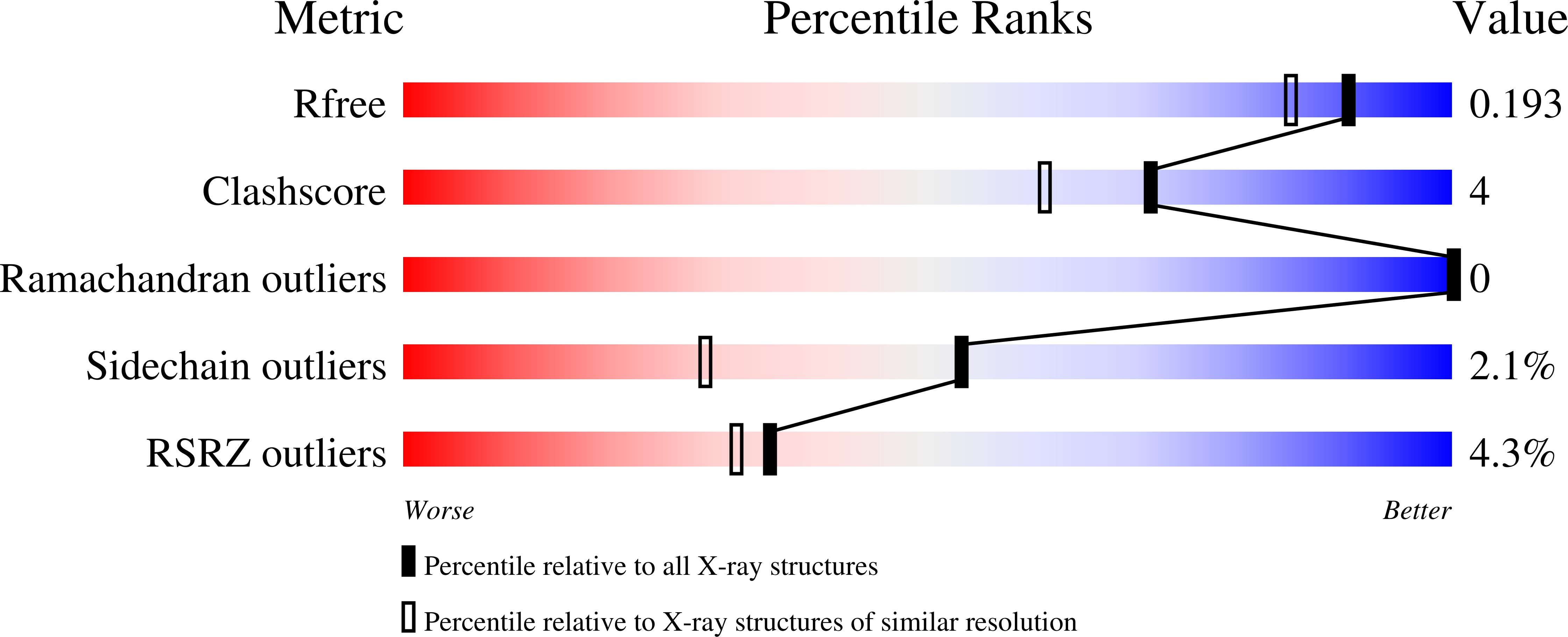
Resolution:
1.60 Å
R-Value Free:
0.18
R-Value Work:
0.15
R-Value Observed:
0.15
Space Group:
C 1 2 1
Macromolecular Entities
Structures with similar UniProt ID
Protein Blast
Polymer Type:polypeptide(L)
Molecule:E3 ubiquitin-protein ligase UHRF1
Gene (Uniprot):Uhrf1
Chain IDs:E (auth: A), F (auth: B)
Chain Length:210
Number of Molecules:2
Biological Source:Mus musculus
Polymer Type:polydeoxyribonucleotide
Molecule:DNA (5'-D(*DCP*DTP*DAP*DCP*DCP*DGP*DGP*DAP*DTP*DTP*DGP*DC)-3')
Chain IDs:A (auth: C), C (auth: E)
Chain Length:12
Number of Molecules:2
Biological Source:
Polymer Type:polydeoxyribonucleotide
Molecule:DNA (5'-D(*DGP*DCP*DAP*DAP*DTP*DCP*(5CM)P*DGP*DGP*DTP*DAP*DG)-3')
Chain IDs:B (auth: D), D (auth: F)
Chain Length:12
Number of Molecules:2
Biological Source:
Modified Residue
Compound ID Chain ID Parent Comp ID Details 2D Image
5CM B DC ?
Primary Citation
Recognition of hemi-methylated DNA by the SRA protein UHRF1 by a base-flipping mechanism
Nature 455 818 821 (2008)
PMID: 18772891 DOI: 10.1038/nature07249

Abstact

DNA methylation of CpG dinucleotides is an important epigenetic modification of mammalian genomes and is essential for the regulation of chromatin structure, of gene expression and of genome stability. Differences in DNA methylation patterns underlie a wide range of biological processes, such as genomic imprinting, inactivation of the X chromosome, embryogenesis, and carcinogenesis. Inheritance of the epigenetic methylation pattern is mediated by the enzyme DNA methyltransferase 1 (Dnmt1), which methylates newly synthesized CpG sequences during DNA replication, depending on the methylation status of the template strands. The protein UHRF1 (also known as Np95 and ICBP90) recognizes hemi-methylation sites via a SET and RING-associated (SRA) domain and directs Dnmt1 to these sites. Here we report the crystal structures of the SRA domain in free and hemi-methylated DNA-bound states. The SRA domain folds into a globular structure with a basic concave surface formed by highly conserved residues. Binding of DNA to the concave surface causes a loop and an amino-terminal tail of the SRA domain to fold into DNA interfaces at the major and minor grooves of the methylation site. In contrast to fully methylated CpG sites recognized by the methyl-CpG-binding domain, the methylcytosine base at the hemi-methylated site is flipped out of the DNA helix in the SRA-DNA complex and fits tightly into a protein pocket on the concave surface. The complex structure suggests that the successive flip out of the pre-existing methylated cytosine and the target cytosine to be methylated is associated with the coordinated transfer of the hemi-methylated CpG site from UHRF1 to Dnmt1.

Legend

Protein

Chemical

Disease

Primary Citation of related structures