2ZJT image
Deposition Date 2008-03-10
Release Date 2009-03-10
Last Version Date 2024-03-13
Entry Detail
PDB ID:
2ZJT
Keywords:
Title:
Crystal structure of dna gyrase B' domain sheds lights on the mechanism for T-segment navigation
Biological Source:
Source Organism:
Host Organism:
Method Details:
Experimental Method:
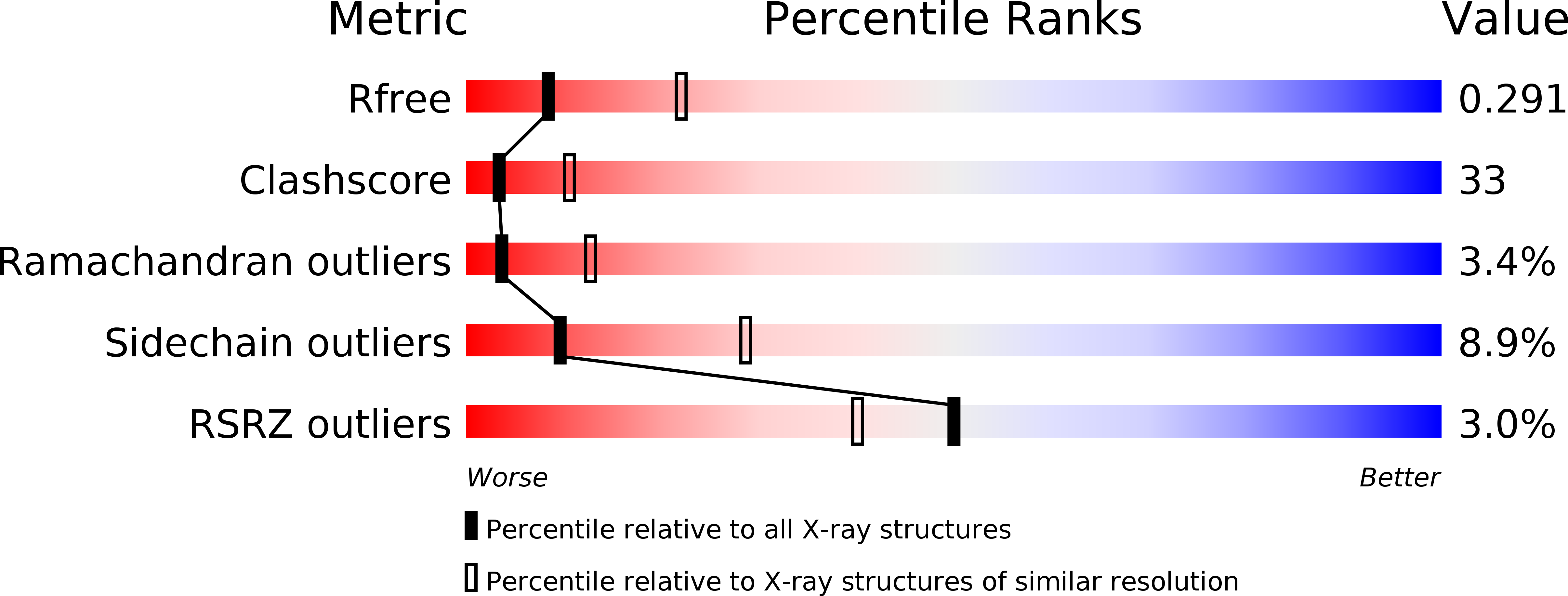
Resolution:
2.80 Å
R-Value Free:
0.28
R-Value Work:
0.24
R-Value Observed:
0.24
Space Group:
P 21 21 21
Macromolecular Entities
Polymer Type:polypeptide(L)
Molecule:DNA gyrase subunit B
Chain IDs:A, B
Chain Length:247
Number of Molecules:2
Biological Source:Mycobacterium tuberculosis
Ligand Molecules
Primary Citation
Crystal structure of DNA gyrase B' domain sheds lights on the mechanism for T-segment navigation
Nucleic Acids Res. 37 5908 5916 (2009)
PMID: 19596812 DOI: 10.1093/nar/gkp586

Abstact

DNA gyrase is an indispensible marvelous molecular machine in manipulating the DNA topology for the prokaryotes. In the 'two-gate' mechanism of DNA topoisomerase, T-segment navigation from N- to DNA-gate is a critical step, but the structural basis supporting this scheme is unclear. The crystal structure of DNA gyrase B' subfragment from Mycobacterium tuberculosis reveals an intrinsic homodimer. The two subunits, each consisting of a Tail and a Toprim domain, are tightly packed one another to form a 'crab-like' organization never observed previously from yeast topo II. Structural comparisons show two orientational alterations of the Tail domain, which may be dominated by a 43-residue peptide at the B' module C-terminus. A highly conserved pentapeptide mediates large-scale intrasubunit conformational change as a hinge point. Mutational studies highlight the significant roles of a negatively charge cluster on a groove at dimer interface. On the basis of structural analysis and mutation experiments, a sluice-like model for T-segment transport is proposed.

Legend

Protein

Chemical

Disease

Primary Citation of related structures