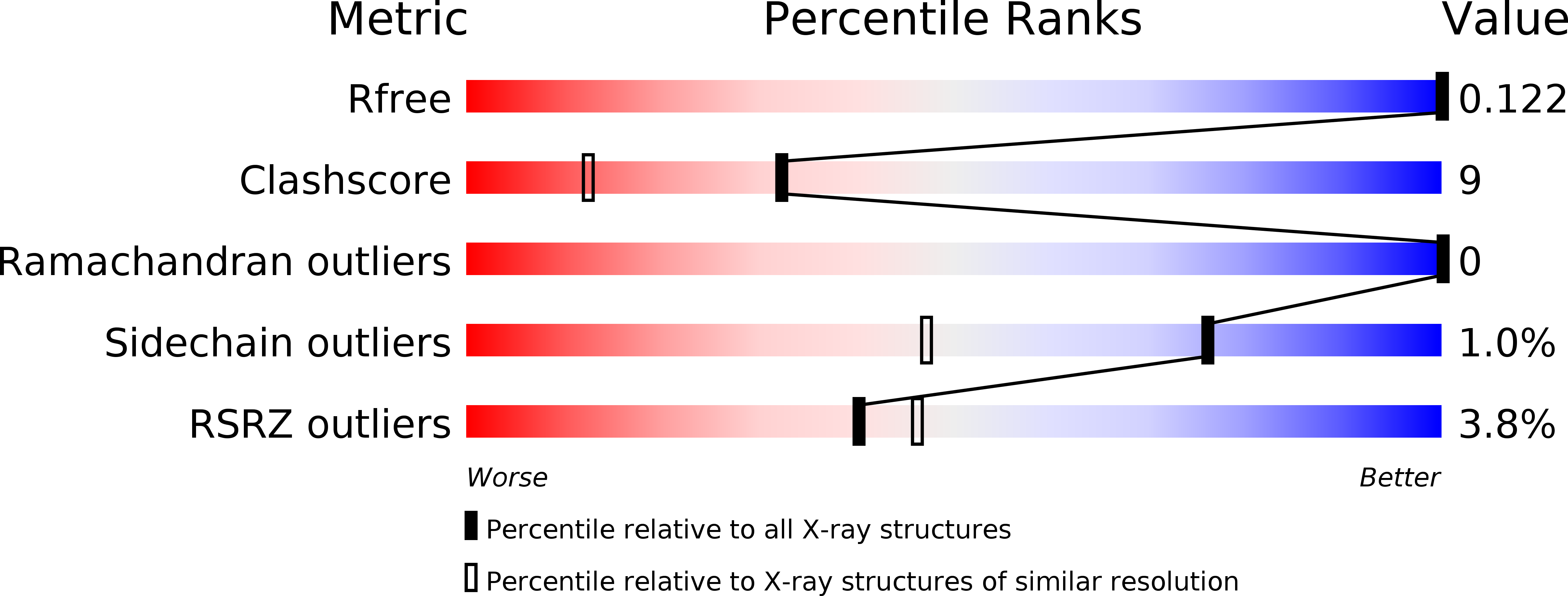
2ZIB image
Deposition Date 2008-02-14
Release Date 2008-08-19
Last Version Date 2024-11-20
Entry Detail
PDB ID:
2ZIB
Title:
Crystal structure analysis of calcium-independent type II antifreeze protein
Biological Source:
Source Organism:
Host Organism:
Method Details:
Experimental Method:
Resolution:
1.34 Å
R-Value Free:
0.16
R-Value Work:
0.13
R-Value Observed:
0.13
Space Group:
P 21 21 21
Macromolecular Entities
Polymer Type:polypeptide(L)
Molecule:Type II antifreeze protein
Chain IDs:A
Chain Length:133
Number of Molecules:1
Biological Source:Brachyopsis rostratus
Ligand Molecules
Primary Citation
Crystal structure and mutational analysis of Ca2+-independent type II antifreeze protein from longsnout poacher, Brachyopsis rostratus
J.Mol.Biol. 382 734 746 (2008)
PMID: 18674542 DOI: 10.1016/j.jmb.2008.07.042

Abstact

We recently found that longsnout poacher (Brachyosis rostratus) produces a Ca(2+)-independent type II antifreeze protein (lpAFP) and succeeded in expressing recombinant lpAFP using Phichia pastoris. Here, we report, for the first time, the X-ray crystal structure of lpAFP at 1.34 A resolution. The lpAFP structure displayed a relatively planar surface, which encompasses two loop regions (Cys86-Lys89 and Asn91-Cys97) and a short beta-strand (Trp109-Leu112) with three unstructured segments (Gly57-Ile58, Ala103-Ala104, and Pro113-His118). Electrostatic calculation of the protein surface showed that the relatively planar surface was divided roughly into a hydrophobic area (composed of the three unstructured segments lacking secondary structure) and a hydrophilic area (composed of the loops and beta-strand). Site-directed mutation of Ile58 with Phe at the center of the hydrophobic area decreased activity significantly, whereas mutation of Leu112 with Phe at an intermediate area between the hydrophobic and hydrophilic areas retained complete activity. In the hydrophilic area, a peptide-swap mutant in the loops retained 60% activity despite simultaneous mutations of eight residues. We conclude that the epicenter of the ice-binding site of lpAFP is the hydrophobic region, which is centered by Ile58, in the relatively planar surface. We built an ice-binding model for lpAFP on the basis of a lattice match of ice and constrained water oxygen atoms surrounding the hydrophobic area in the lpAFP structure. The model in which lpAFP has been docked to a secondary prism (2-1-10) plane, which is different from the one determined for Ca(2+)-independent type II AFP from sea raven (11-21), appears to explain the results of the mutagenesis analysis.

Legend

Protein

Chemical

Disease

Primary Citation of related structures