Deposition Date
2008-02-11
Release Date
2008-05-20
Last Version Date
2023-11-01
Entry Detail
PDB ID:
2ZHX
Keywords:
Title:
Crystal structure of Uracil-DNA Glycosylase from Mycobacterium tuberculosis in complex with a proteinaceous inhibitor
Biological Source:
Source Organism(s):
Mycobacterium tuberculosis H37Rv (Taxon ID: 83332)
Bacillus phage PBS2 (Taxon ID: 10684)
Bacillus phage PBS2 (Taxon ID: 10684)
Expression System(s):
Method Details:
Experimental Method:
Resolution:
3.10 Å
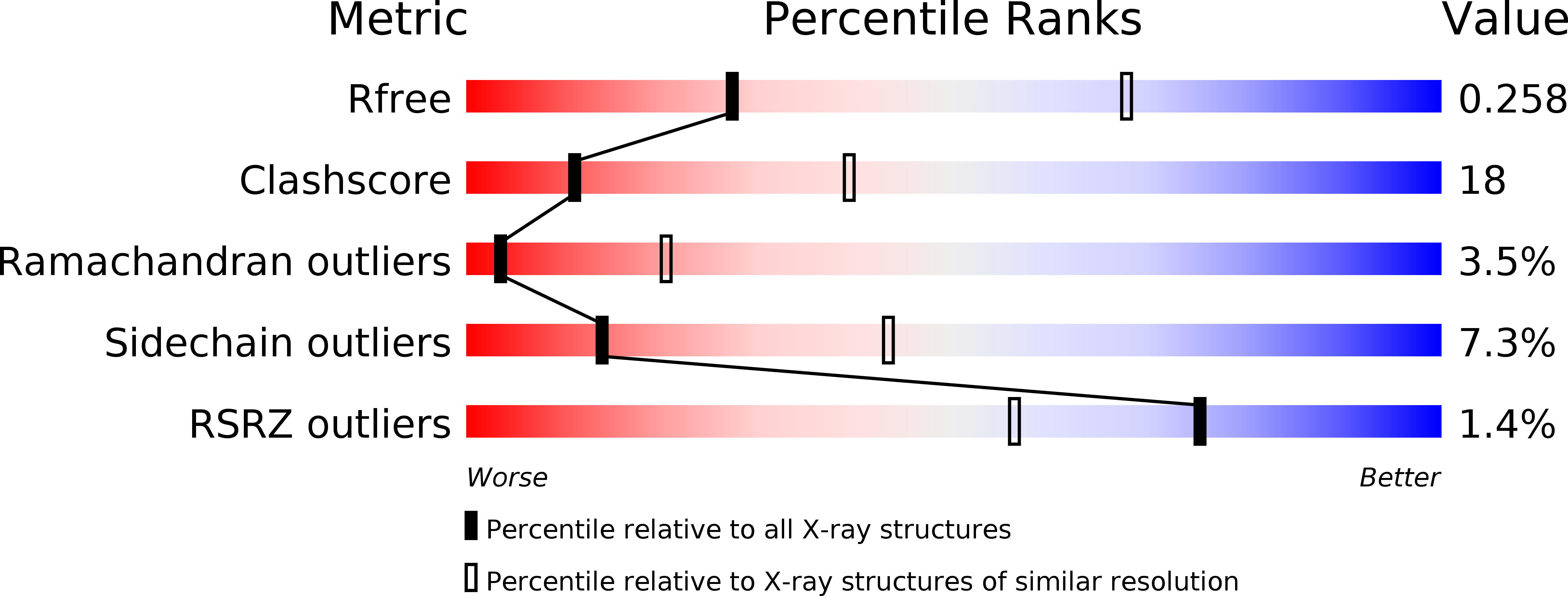
R-Value Free:
0.27
R-Value Work:
0.23
R-Value Observed:
0.23
Space Group:
C 1 2 1