Deposition Date
2008-01-07
Release Date
2008-09-23
Last Version Date
2023-11-01
Entry Detail
PDB ID:
2ZFI
Keywords:
Title:
Crystal Structure of the Kif1A Motor Domain Before Mg Release
Biological Source:
Source Organism(s):
Mus musculus (Taxon ID: 10090)
Expression System(s):
Method Details:
Experimental Method:
Resolution:
1.55 Å
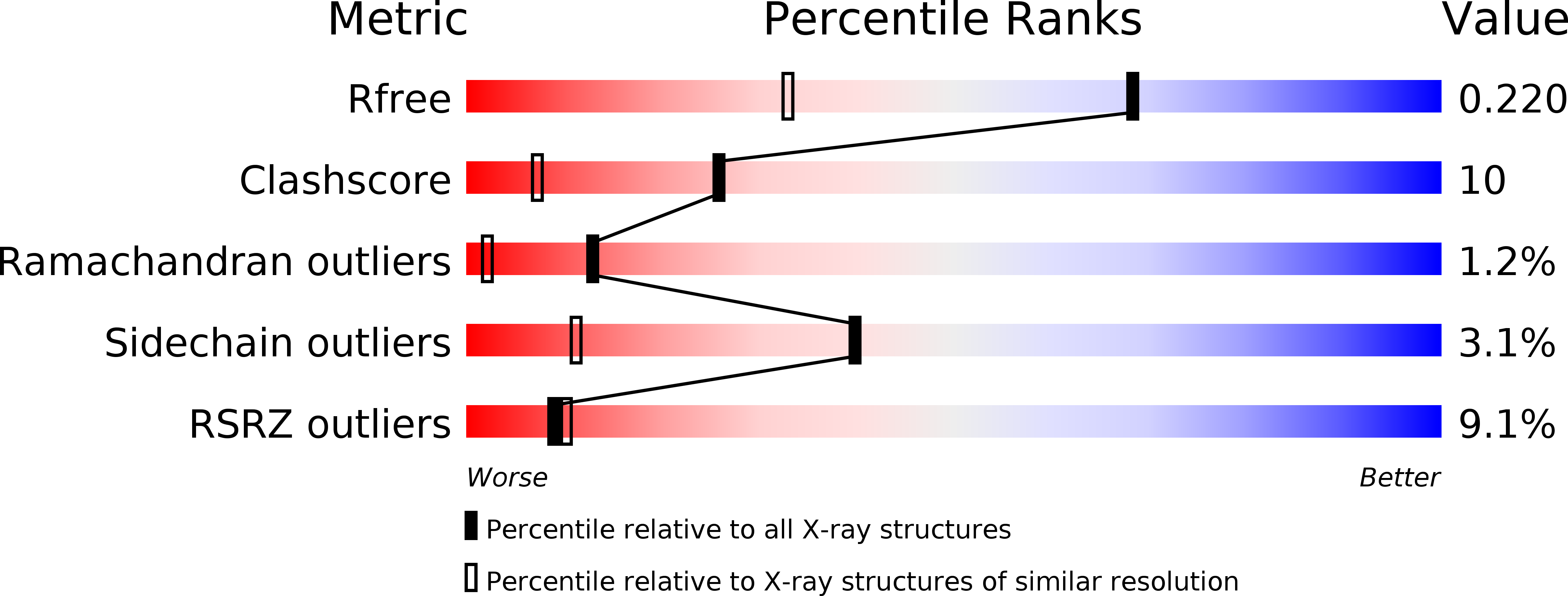
R-Value Free:
0.22
R-Value Work:
0.19
R-Value Observed:
0.19
Space Group:
P 21 21 21