2ZFD image
Deposition Date 2007-12-29
Release Date 2008-02-19
Last Version Date 2024-03-13
Entry Detail
PDB ID:
2ZFD
Title:
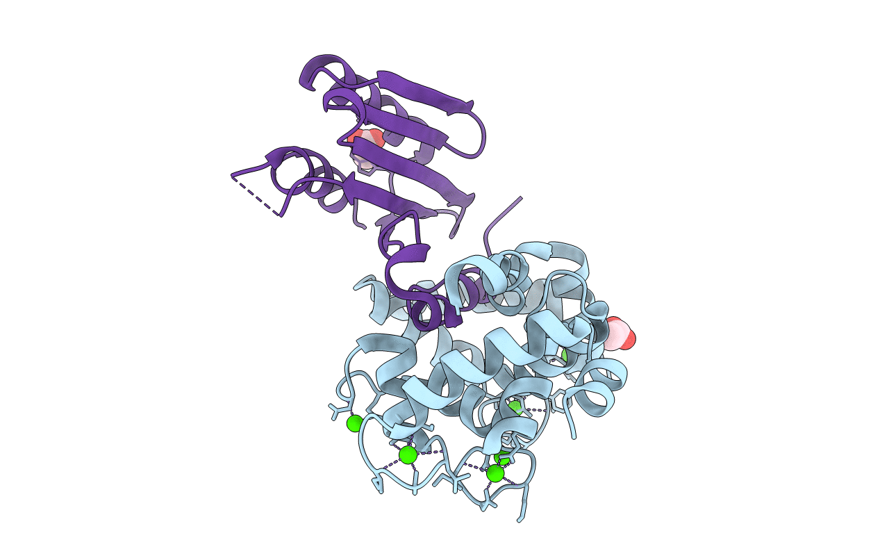
The crystal structure of plant specific calcium binding protein AtCBL2 in complex with the regulatory domain of AtCIPK14
Biological Source:
Source Organism:
Host Organism:
Method Details:
Experimental Method:
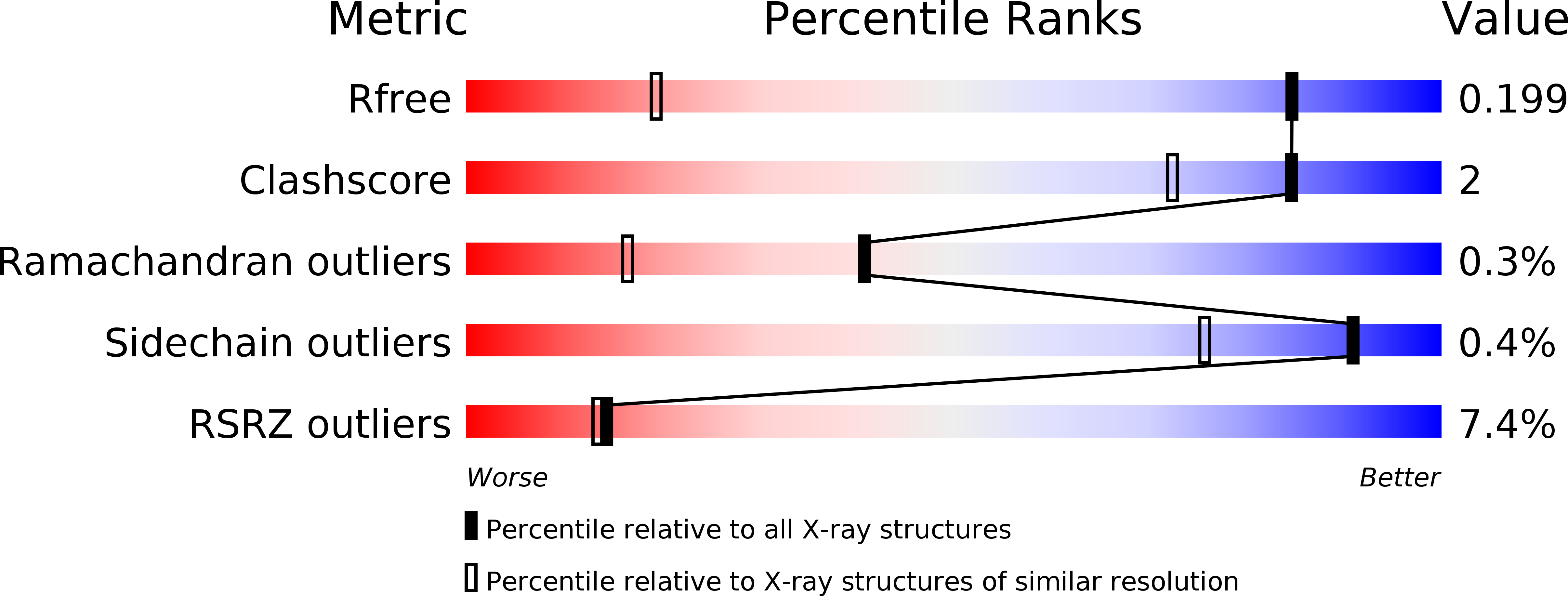
Resolution:
1.20 Å
R-Value Free:
0.19
R-Value Work:
0.17
R-Value Observed:
0.18
Space Group:
P 1 21 1
Macromolecular Entities
Structures with similar UniProt ID
Protein Blast
Polymer Type:polypeptide(L)
Molecule:Calcineurin B-like protein 2
Gene (Uniprot):CBL2
Chain IDs:A
Chain Length:226
Number of Molecules:1
Biological Source:Arabidopsis thaliana
Structures with similar UniProt ID
Protein Blast
Polymer Type:polypeptide(L)
Molecule:Putative uncharacterized protein T20L15_90
Gene (Uniprot):CIPK14
Chain IDs:B
Chain Length:123
Number of Molecules:1
Biological Source:Arabidopsis thaliana
Primary Citation
The crystal structure of plant-specific calcium-binding protein AtCBL2 in complex with the regulatory domain of AtCIPK14
J.Mol.Biol. 377 246 257 (2008)
PMID: 18237745 DOI: 10.1016/j.jmb.2008.01.006

Abstact

Calcium signals mediate a multitude of plant responses to external stimuli. Calcineurin B-like (CBL) proteins and their target kinases, CBL-interacting protein kinases (CIPKs), represent important relays in plant calcium signaling. CBL interacts with CIPK through a conserved motif (NAF/FISL motif) within the C-terminal regulatory domain. To better understand the functional role of the CBL-CIPK system, we determined the crystal structure of AtCBL2 in complex with the regulatory domain of AtCIPK14 at 1.2 A resolution. The NAF/FISL motif is inserted into a hydrophobic crevice within AtCBL2, accompanied by a large displacement of the helices and loop on the opposite side of the NAF/FISL motif from the C-terminal region, which shields the hydrophobic crevice in free form. Ca(2+) are coordinated within four EF hands in AtCBL2 in bound form. This calcium coordination pattern differs from that in the structure of the SOS3-SOS2 complex previously reported. Structural comparison of the two structures shows that the recognition of CBL by CIPK is performed in a similar manner, but inherent interactions confer binding affinity and specificity.

Legend

Protein

Chemical

Disease

Primary Citation of related structures