Deposition Date
2007-12-26
Release Date
2008-12-30
Last Version Date
2024-10-30
Entry Detail
PDB ID:
2ZF9
Keywords:
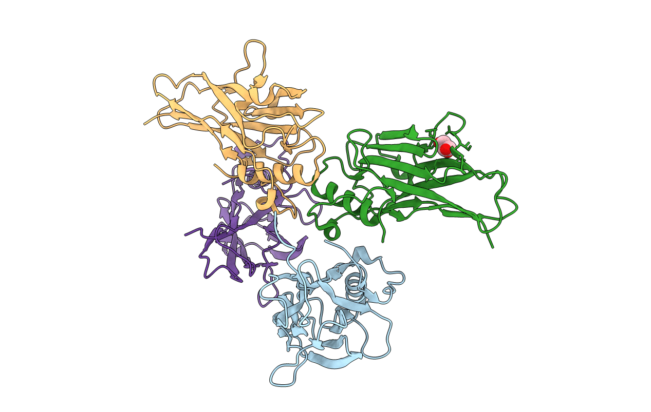
Title:
Crystal structure of a type III cohesin module from the cellulosomal ScaE cell-surface anchoring scaffoldin of Ruminococcus flavefaciens
Biological Source:
Source Organism(s):
Ruminococcus flavefaciens (Taxon ID: 1265)
Expression System(s):
Method Details:
Experimental Method:
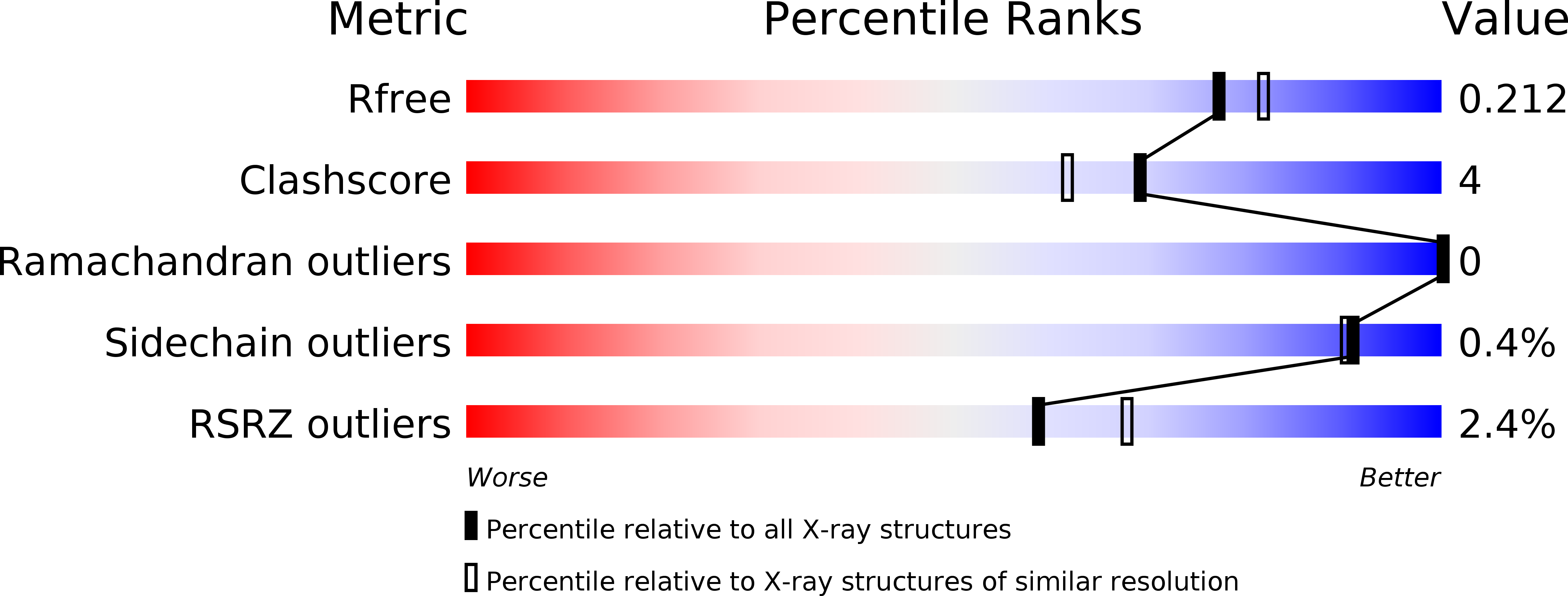
Resolution:
1.95 Å
R-Value Free:
0.21
R-Value Work:
0.16
R-Value Observed:
0.16
Space Group:
C 1 2 1