Deposition Date
2007-12-20
Release Date
2008-05-06
Last Version Date
2023-11-01
Entry Detail
PDB ID:
2ZF5
Keywords:
Title:
Crystal Structure of highly thermostable glycerol kinase from a hyperthermophilic archaeon
Biological Source:
Source Organism(s):
Thermococcus kodakarensis (Taxon ID: 69014)
Expression System(s):
Method Details:
Experimental Method:
Resolution:
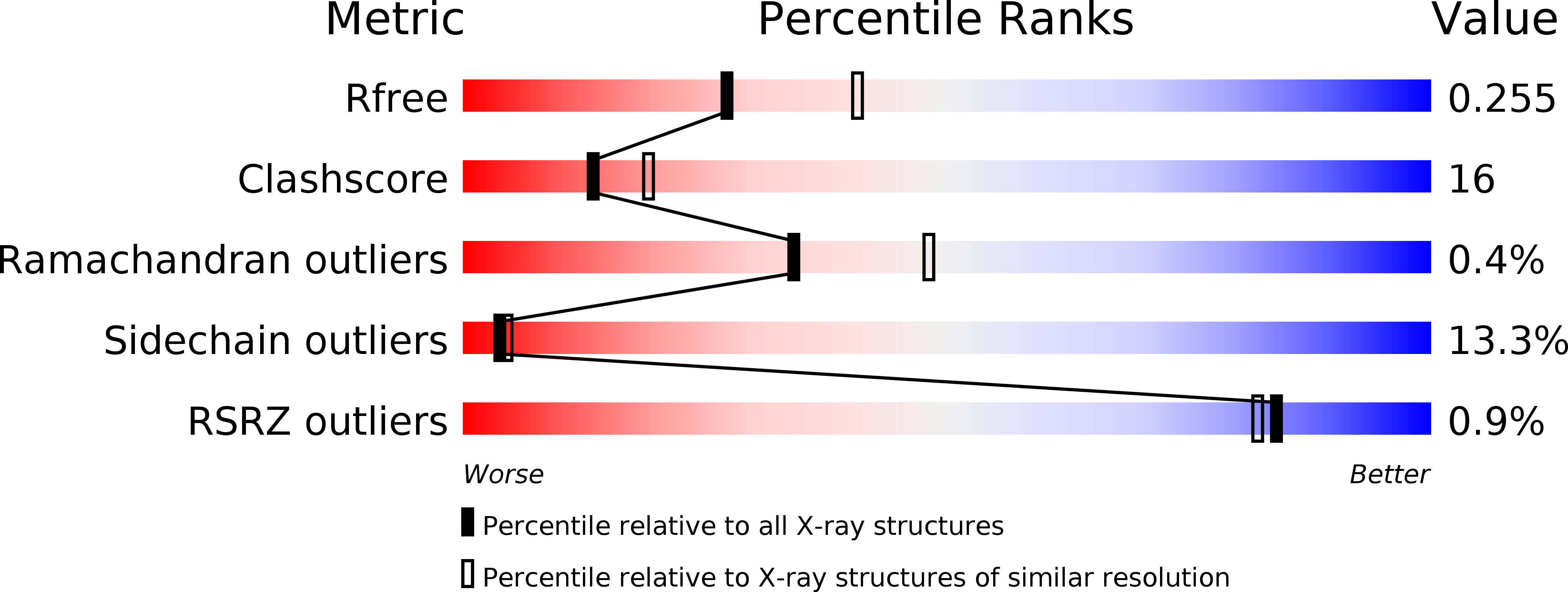
2.40 Å
R-Value Free:
0.25
R-Value Work:
0.17
R-Value Observed:
0.17
Space Group:
H 3