Deposition Date
2007-12-12
Release Date
2008-04-22
Last Version Date
2023-11-01
Entry Detail
PDB ID:
2ZED
Keywords:
Title:
Crystal structure of the human glutaminyl cyclase mutant S160A at 1.7 angstrom resolution
Biological Source:
Source Organism(s):
Homo sapiens (Taxon ID: 9606)
Expression System(s):
Method Details:
Experimental Method:
Resolution:
1.70 Å
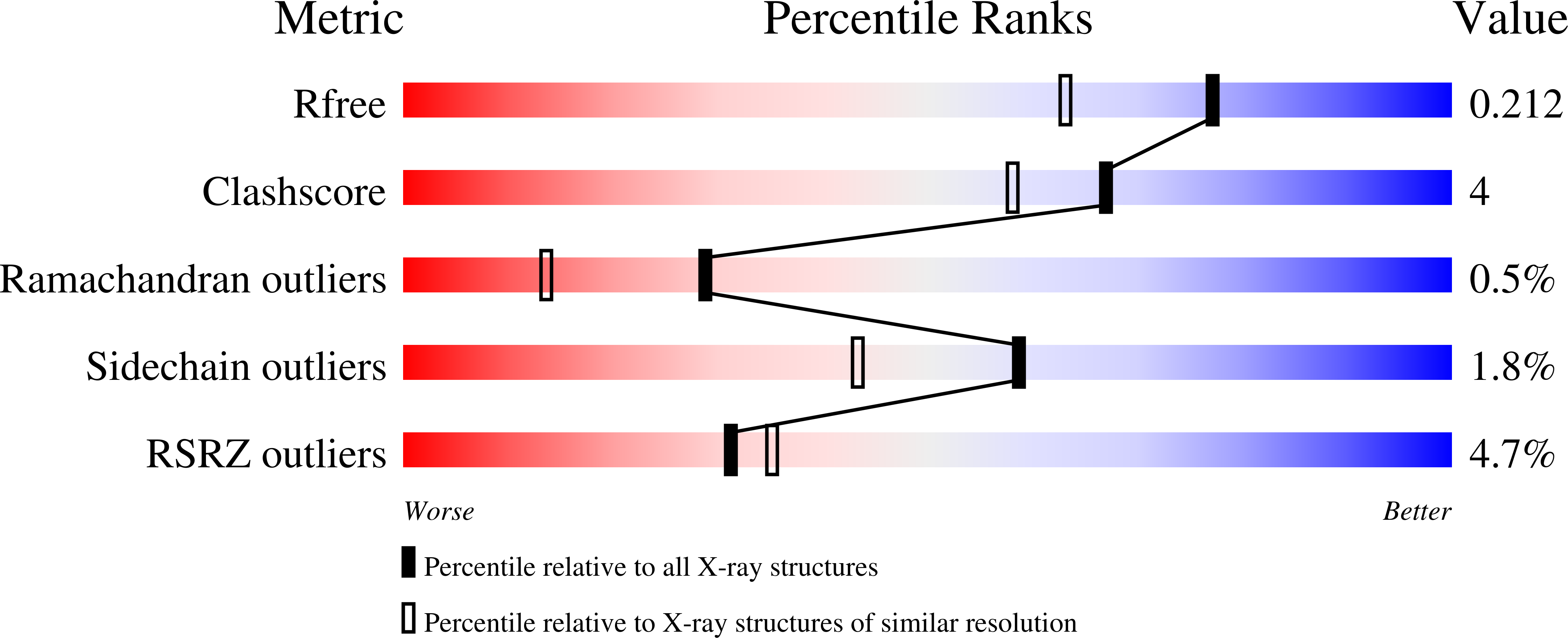
R-Value Free:
0.21
R-Value Work:
0.18
R-Value Observed:
0.18
Space Group:
H 3 2