
Deposition Date
2007-11-09
Release Date
2008-04-15
Last Version Date
2023-08-30
Entry Detail
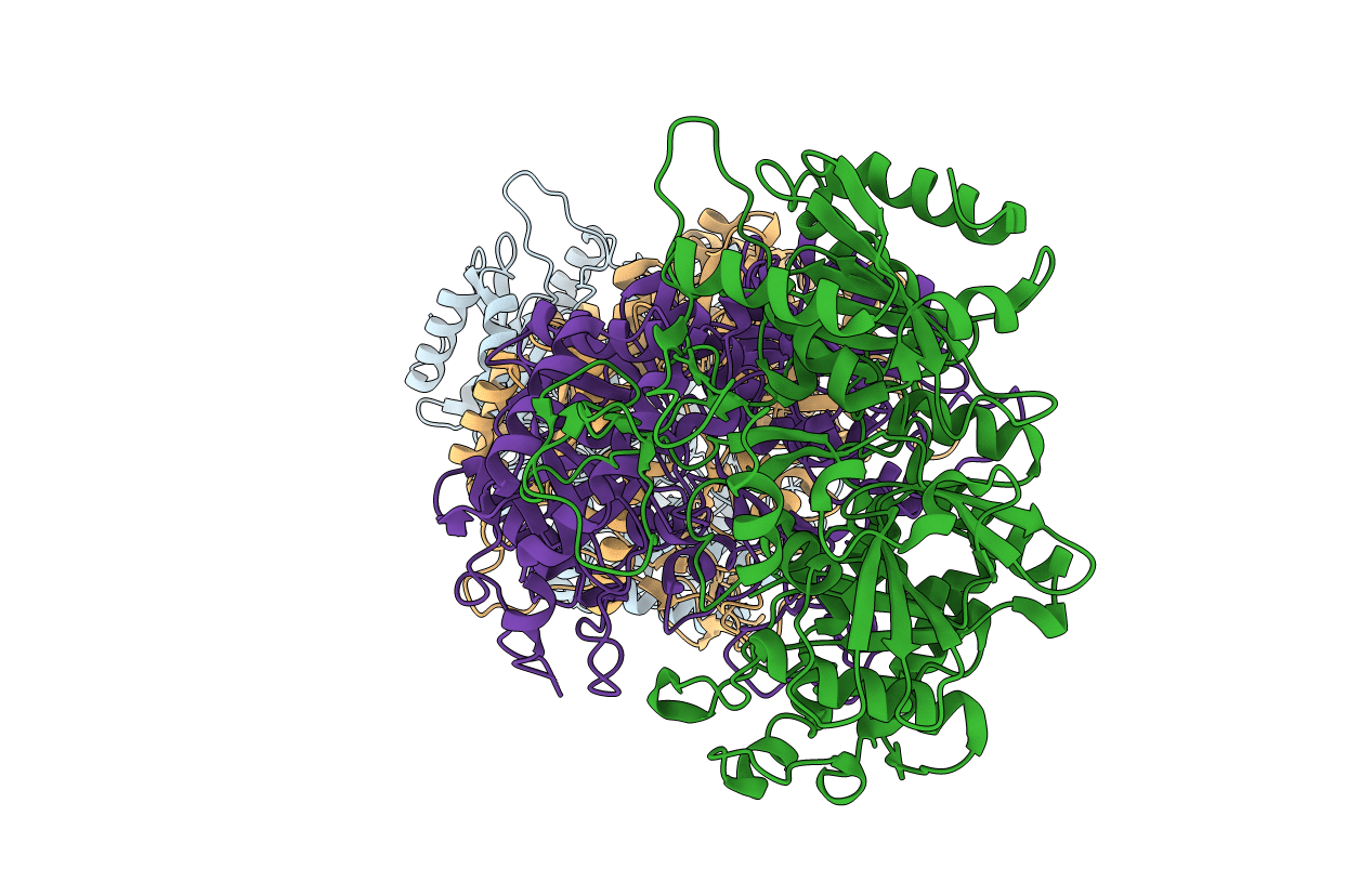
PDB ID:
2ZCI
Keywords:
Title:
Structure of a GTP-dependent bacterial PEP-carboxykinase from Corynebacterium glutamicum
Biological Source:
Source Organism(s):
Corynebacterium glutamicum (Taxon ID: 1718)
Method Details:
Experimental Method:
Resolution:
2.30 Å
R-Value Free:
0.27
R-Value Work:
0.18
R-Value Observed:
0.19
Space Group:
P 1 21 1
