Deposition Date
2007-11-06
Release Date
2008-10-28
Last Version Date
2024-10-30
Entry Detail
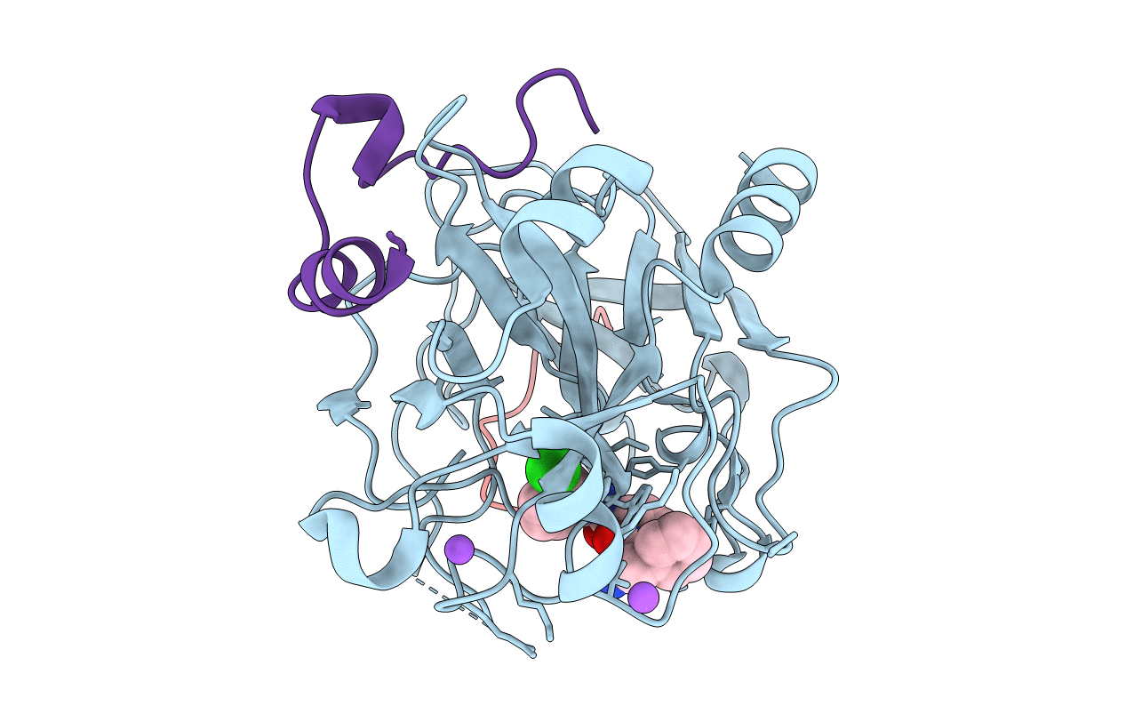
Biological Source:
Source Organism(s):
Hirudo medicinalis (Taxon ID: 6421)
Homo sapiens (Taxon ID: 9606)
Homo sapiens (Taxon ID: 9606)
Method Details:
Experimental Method:
Resolution:
1.58 Å
R-Value Free:
0.23
R-Value Work:
0.18
R-Value Observed:
0.18
Space Group:
C 1 2 1