2ZBZ image
Entry Detail
PDB ID:
2ZBZ
Keywords:
Title:
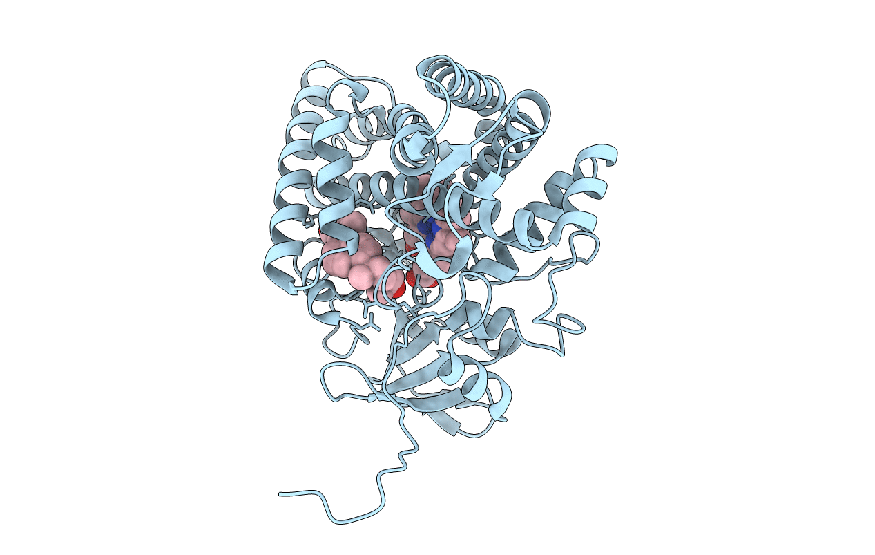
Crystal structure of vitamin D hydroxylase cytochrome P450 105A1 (R84A mutant) in complex with 1,25-dihydroxyvitamin D3
Biological Source:
Source Organism:
Host Organism:
PDB Version:
Deposition Date:
2007-10-30
Release Date:
2008-04-08
Method Details:
Experimental Method:
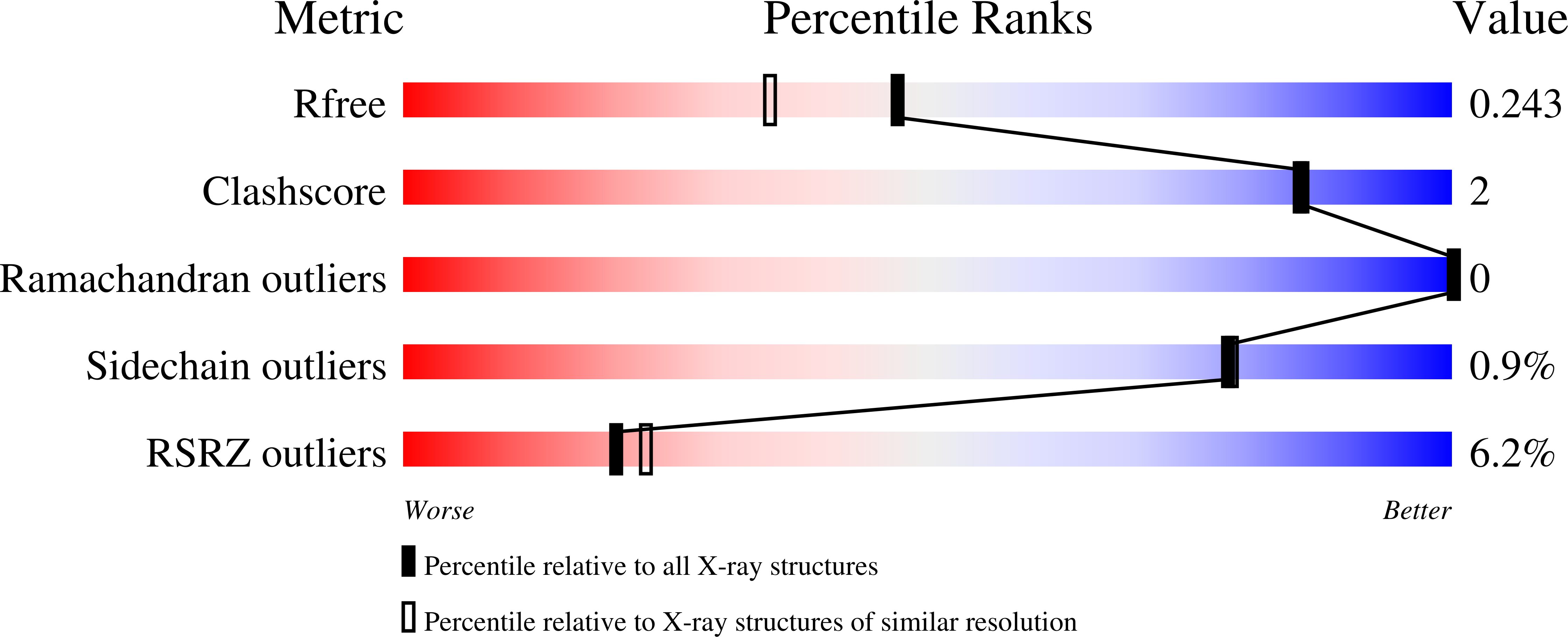
Resolution:
1.90 Å
R-Value Free:
0.24
R-Value Work:
0.19
R-Value Observed:
0.20
Space Group:
P 21 21 21
Macromolecular Entities
Polymer Type:polypeptide(L)
Description:Cytochrome P450-SU1
Mutations:R84A
Chain IDs:A
Chain Length:412
Number of Molecules:1
Biological Source:Streptomyces griseolus
Primary Citation
Crystal Structure of CYP105A1 (P450SU-1) in Complex with 1alpha,25-Dihydroxyvitamin D3
Biochemistry 47 4017 4027 (2008)
PMID: 18314962 DOI: 10.1021/bi7023767

Abstact

Vitamin D 3 (VD 3), a prohormone in mammals, plays a crucial role in the maintenance of calcium and phosphorus concentrations in serum. Activation of VD 3 requires 25-hydroxylation in the liver and 1alpha-hydroxylation in the kidney by cytochrome P450 (CYP) enzymes. Bacterial CYP105A1 converts VD 3 into 1alpha,25-dihydroxyvitamin D 3 (1alpha,25(OH) 2D 3) in two independent reactions, despite its low sequence identity with mammalian enzymes (<21% identity). The present study determined the crystal structures of a highly active mutant (R84A) of CYP105A1 from Streptomyces griseolus in complex and not in complex with 1alpha,25(OH) 2D 3. The compound 1alpha,25(OH) 2D 3 is positioned 11 A from the iron atom along the I helix within the pocket. A similar binding mode is observed in the structure of the human CYP2R1-VD 3 complex, indicating a common substrate-binding mechanism for 25-hydroxylation. A comparison with the structure of wild-type CYP105A1 suggests that the loss of two hydrogen bonds in the R84A mutant increases the adaptability of the B' and F helices, creating a transient binding site. Further mutational analysis of the active site reveals that 25- and 1alpha-hydroxylations share residues that participate in these reactions. These results provide the structural basis for understanding the mechanism of the two-step hydroxylation that activates VD 3.

Legend

Protein

Chemical

Disease

Primary Citation of related structures