Deposition Date
2007-10-16
Release Date
2008-09-30
Last Version Date
2023-11-01
Entry Detail
PDB ID:
2ZB3
Keywords:
Title:
Crystal structure of mouse 15-ketoprostaglandin delta-13-reductase in complex with NADPH
Biological Source:
Source Organism(s):
Mus musculus (Taxon ID: 10090)
Expression System(s):
Method Details:
Experimental Method:
Resolution:
2.00 Å
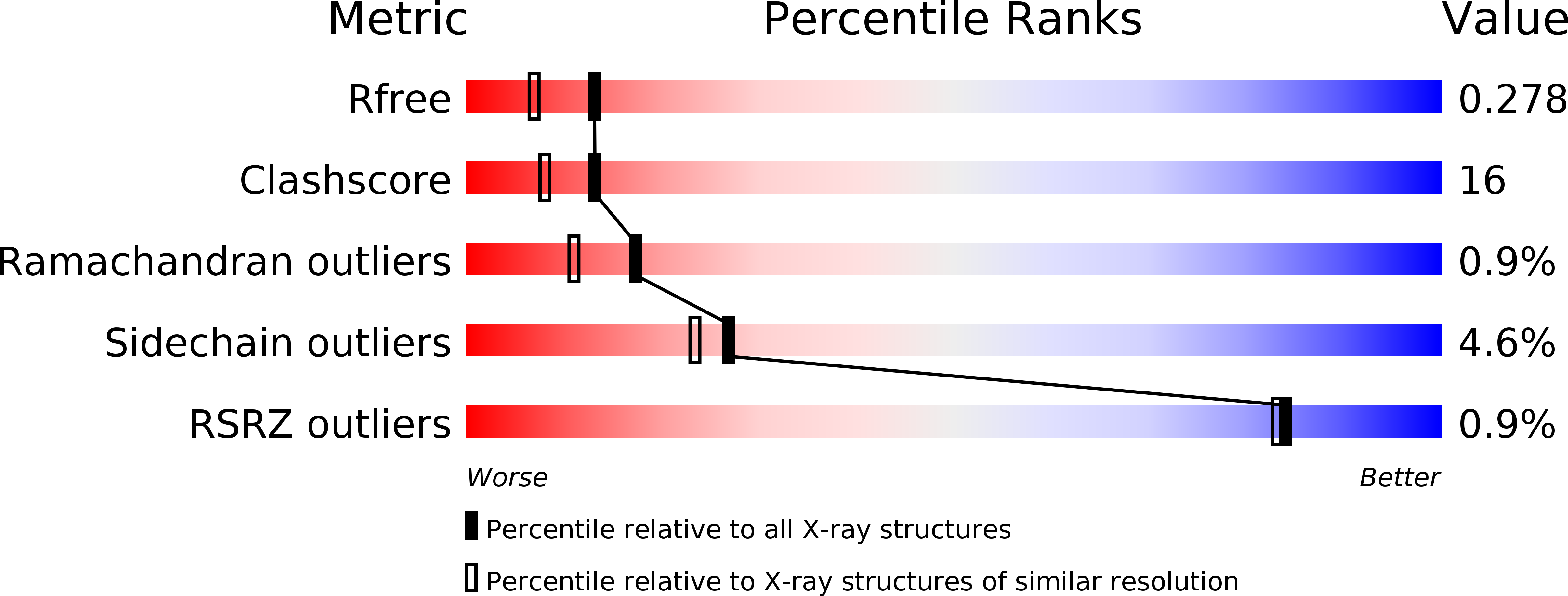
R-Value Free:
0.27
R-Value Work:
0.20
R-Value Observed:
0.21
Space Group:
P 41 21 2