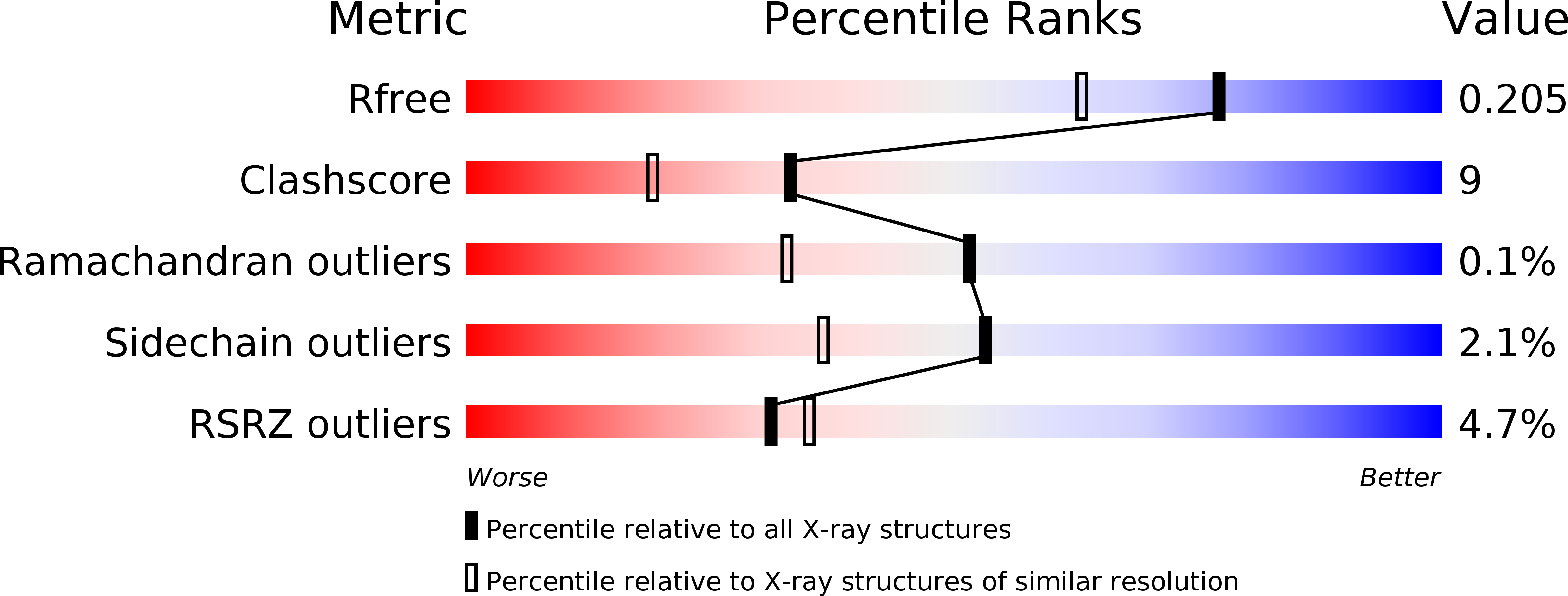
2Z8G image
Deposition Date 2007-09-05
Release Date 2007-12-18
Last Version Date 2024-10-16
Entry Detail
PDB ID:
2Z8G
Keywords:
Title:
Aspergillus niger ATCC9642 isopullulanase complexed with isopanose
Biological Source:
Source Organism:
Host Organism:
Method Details:
Experimental Method:
Resolution:
1.70 Å
R-Value Free:
0.21
R-Value Work:
0.18
R-Value Observed:
0.18
Space Group:
P 21 21 21
Macromolecular Entities
Polymer Type:polypeptide(L)
Molecule:Isopullulanase
Gene (Uniprot):ipuA
Chain IDs:A, B
Chain Length:549
Number of Molecules:2
Biological Source:Aspergillus niger
Modified Residue
Compound ID Chain ID Parent Comp ID Details 2D Image
ASN A ASN GLYCOSYLATION SITE
Ligand Molecules
Primary Citation
Crystal Structure of Aspergillus niger Isopullulanase, a Member of Glycoside Hydrolase Family 49
J.Mol.Biol. 376 210 220 (2008)
PMID: 18155243 DOI: 10.1016/j.jmb.2007.11.098

Abstact

An isopullulanase (IPU) from Aspergillus niger ATCC9642 hydrolyzes alpha-1,4-glucosidic linkages of pullulan to produce isopanose. Although IPU does not hydrolyze dextran, it is classified into glycoside hydrolase family 49 (GH49), major members of which are dextran-hydrolyzing enzymes. IPU is highly glycosylated, making it difficult to obtain its crystal. We used endoglycosidase H(f) to cleave the N-linked oligosaccharides of IPU, and we here determined the unliganded and isopanose-complexed forms of IPU, both solved at 1.7-A resolution. IPU is composed of domains N and C joined by a short linker, with electron density maps for 11 or 12 N-acetylglucosamine residues per molecule. Domain N consists of 13 beta-strands and forms a beta-sandwich. Domain C, where the active site is located, forms a right-handed beta-helix, and the lengths of the pitches of each coil of the beta-helix are similar to those of GH49 dextranase and GH28 polygalacturonase. The entire structure of IPU resembles that of a GH49 enzyme, Penicillium minioluteum dextranase (Dex49A), despite a difference in substrate specificity. Compared with the active sites of IPU and Dex49A, the amino acid residues participating in subsites +2 and +3 are not conserved, and the glucose residues of isopanose bound to IPU completely differ in orientation from the corresponding glucose residues of isomaltose bound to Dex49A. The shape of the catalytic cleft characterized by the seventh coil of the beta-helix and a loop from domain N appears to be critical in determining the specificity of IPU for pullulan.

Legend

Protein

Chemical

Disease

Primary Citation of related structures