2Z87 image
Deposition Date 2007-09-03
Release Date 2008-09-16
Last Version Date 2024-03-13
Entry Detail
PDB ID:
2Z87
Keywords:
Title:
Crystal structure of chondroitin polymerase from Escherichia coli strain K4 (K4CP) complexed with UDP-GalNAc and UDP
Biological Source:
Source Organism:
Escherichia coli (Taxon ID: 562)
Host Organism:
Method Details:
Experimental Method:
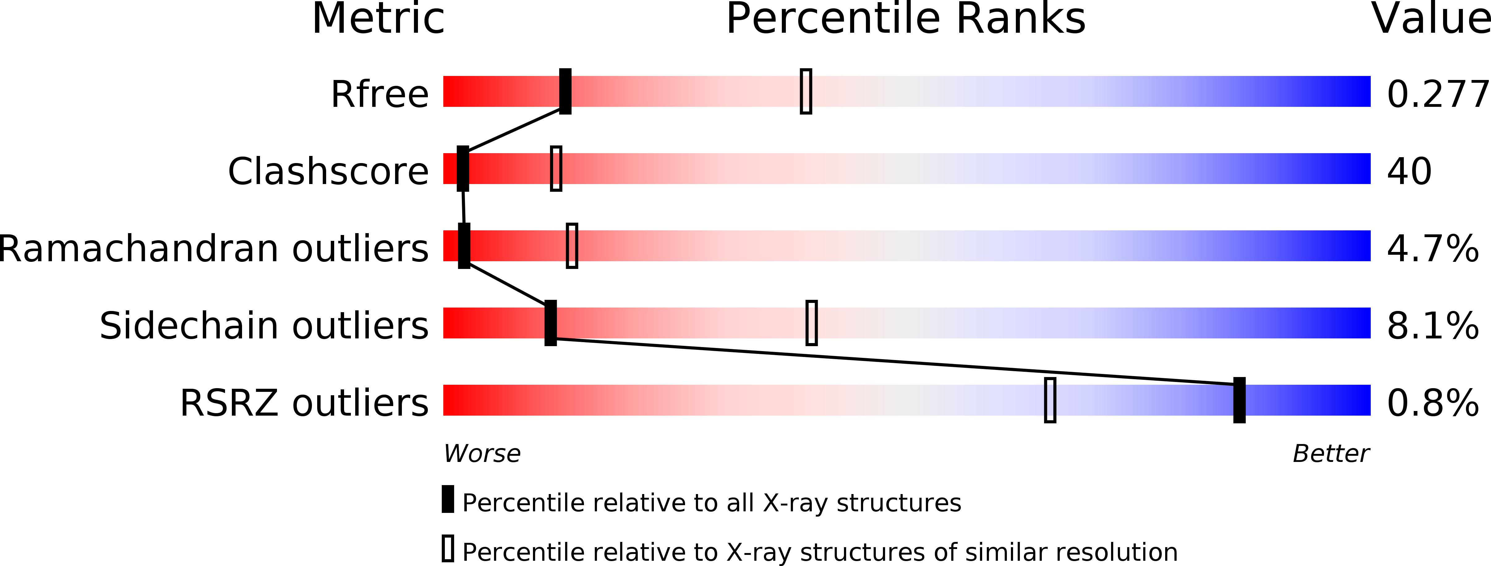
Resolution:
3.00 Å
R-Value Free:
0.28
R-Value Work:
0.19
R-Value Observed:
0.19
Space Group:
P 1 21 1
Macromolecular Entities
Polymer Type:polypeptide(L)
Molecule:Chondroitin synthase
Gene (Uniprot):kfoC
Chain IDs:A, B
Chain Length:624
Number of Molecules:2
Biological Source:Escherichia coli
Primary Citation
Crystal structure of chondroitin polymerase from Escherichia coli K4.
Biochem. Biophys. Res. Commun. 378 10 14 (2009)
PMID: 18771653 DOI: 10.1016/j.bbrc.2008.08.121

Abstact

Elongation of glycosaminoglycan chains, such as heparan and chondroitin, is catalyzed by bi-functional glycosyltransferases, for which both 3-dimensional structures and reaction mechanisms remain unknown. The bacterial chondroitin polymerase K4CP catalyzes elongation of the chondroitin chain by alternatively transferring the GlcUA and GalNAc moiety from UDP-GlcUA and UDP-GalNAc to the non-reducing ends of the chondroitin chain. Here, we have determined the crystal structure of K4CP in the presence of UDP and UDP-GalNAc as well as with UDP and UDP-GlcUA. The structures consisted of two GT-A fold domains in which the two active sites were 60A apart. UDP-GalNAc and UDP-GlcUA were found at the active sites of the N-terminal and C-terminal domains, respectively. The present K4CP structures have provided the structural basis for further investigating the molecular mechanism of biosynthesis of chondroitin chain.

Legend

Protein

Chemical

Disease

Primary Citation of related structures