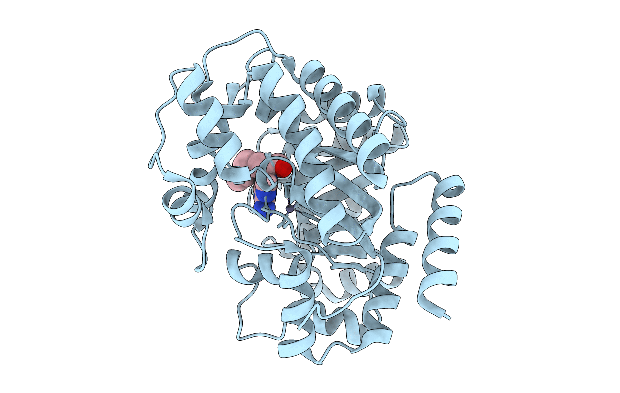
2Z7G image
Deposition Date 2007-08-20
Release Date 2008-07-01
Last Version Date 2023-11-01
Entry Detail
PDB ID:
2Z7G
Keywords:
Title:
Crystal structure of adenosine deaminase ligated with EHNA
Biological Source:
Source Organism:
Bos taurus (Taxon ID: 9913)
Method Details:
Experimental Method:
Resolution:
2.52 Å
R-Value Free:
0.24
R-Value Work:
0.20
R-Value Observed:
0.21
Space Group:
P 43 21 2
Macromolecular Entities
Structures with similar UniProt ID
Protein Blast
Polymer Type:polypeptide(L)
Molecule:Adenosine deaminase
Gene (Uniprot):ADA
Chain IDs:A
Chain Length:356
Number of Molecules:1
Biological Source:Bos taurus
Primary Citation
Conformational change of adenosine deaminase during ligand-exchange in a crystal
Biochem.Biophys.Res.Commun. 373 53 57 (2008)
PMID: 18549808 DOI: 10.1016/j.bbrc.2008.05.180

Abstact

Adenosine deaminase (ADA) perpetuates chronic inflammation by degrading extracellular adenosine which is toxic for lymphocytes. ADA has two distinct conformations: open form and closed form. From the crystal structures with various ligands, the non-nucleoside type inhibitors bind to the active site occupying the critical water-binding-position and sustain the open form of apo-ADA. In contrast, substrate mimics do not occupy the critical position, and induce the large conformational change to the closed form. However, it is difficult to predict the binding of (+)-erythro-9-(2-hydroxy-3-nonyl)adenine (EHNA), as it possesses characteristic parts of both the substrate and the non-nucleoside inhibitors. The crystal structure shows that EHNA binds to the open form through a novel recognition of the adenine base accompanying conformational change from the closed form of the PR-ADA complex in crystalline state.

Legend

Protein

Chemical

Disease

Primary Citation of related structures