2Z73 image
Deposition Date 2007-08-13
Release Date 2008-05-13
Last Version Date 2024-10-16
Entry Detail
PDB ID:
2Z73
Title:
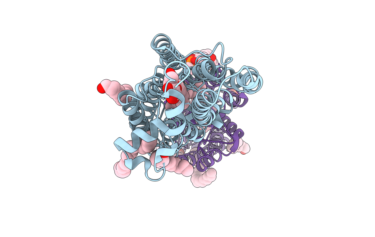
Crystal structure of squid rhodopsin
Biological Source:
Source Organism:
Todarodes pacificus (Taxon ID: )
Method Details:
Experimental Method:
Resolution:
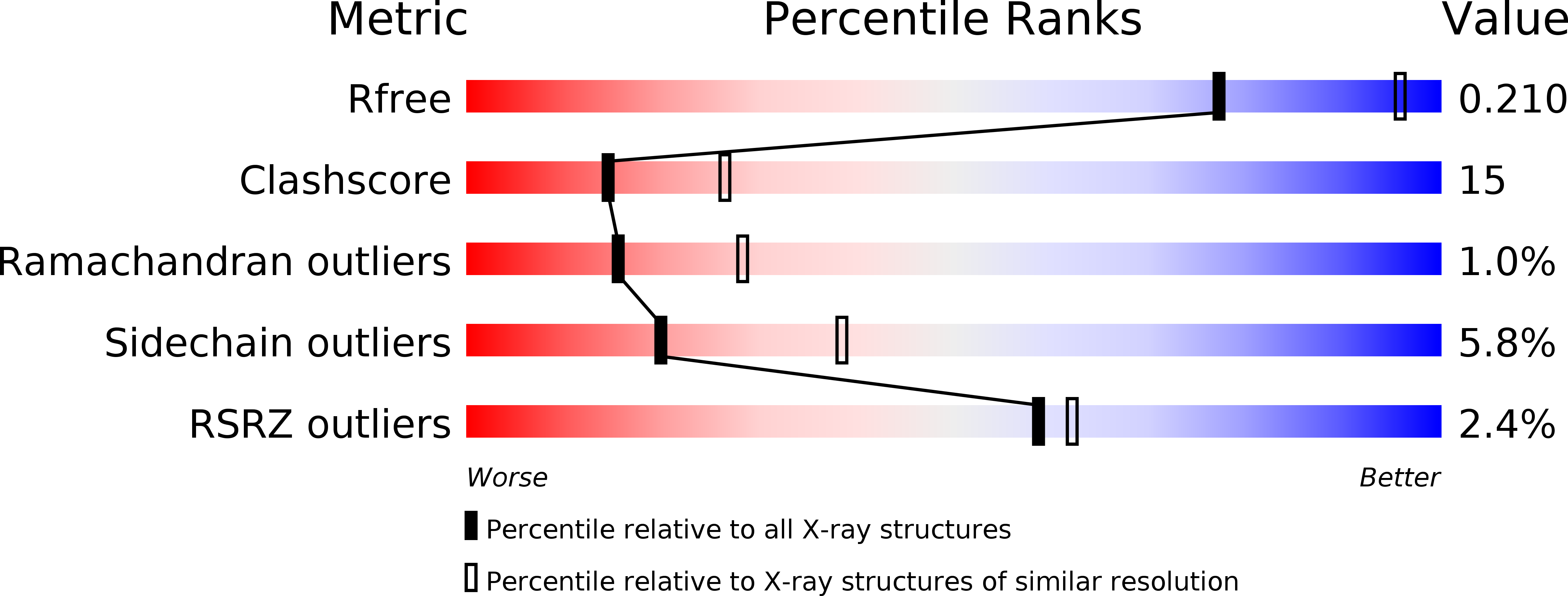
2.50 Å
R-Value Free:
0.20
R-Value Work:
0.18
R-Value Observed:
0.18
Space Group:
P 62
Macromolecular Entities
Structures with similar UniProt ID
Protein Blast
Polymer Type:polypeptide(L)
Molecule:Rhodopsin
Gene (Uniprot):RHO
Chain IDs:A, B
Chain Length:448
Number of Molecules:2
Biological Source:Todarodes pacificus
Primary Citation
Crystal structure of squid rhodopsin.
Nature 453 363 367 (2008)
PMID: 18480818 DOI: 10.1038/nature06925

Abstact

Invertebrate phototransduction uses an inositol-1,4,5-trisphosphate signalling cascade in which photoactivated rhodopsin stimulates a G(q)-type G protein, that is, a class of G protein that stimulates membrane-bound phospholipase Cbeta. The same cascade is used by many G-protein-coupled receptors, indicating that invertebrate rhodopsin is a prototypical member. Here we report the crystal structure of squid (Todarodes pacificus) rhodopsin at 2.5 A resolution. Among seven transmembrane alpha-helices, helices V and VI extend into the cytoplasmic medium and, together with two cytoplasmic helices, they form a rigid protrusion from the membrane surface. This peculiar structure, which is not seen in bovine rhodopsin, seems to be crucial for the recognition of G(q)-type G proteins. The retinal Schiff base forms a hydrogen bond to Asn 87 or Tyr 111; it is far from the putative counterion Glu 180. In the crystal, a tight association is formed between the amino-terminal polypeptides of neighbouring monomers; this intermembrane dimerization may be responsible for the organization of hexagonally packed microvillar membranes in the photoreceptor rhabdom.

Legend

Protein

Chemical

Disease

Primary Citation of related structures
Feedback Form
Name
Email
Institute
Feedback