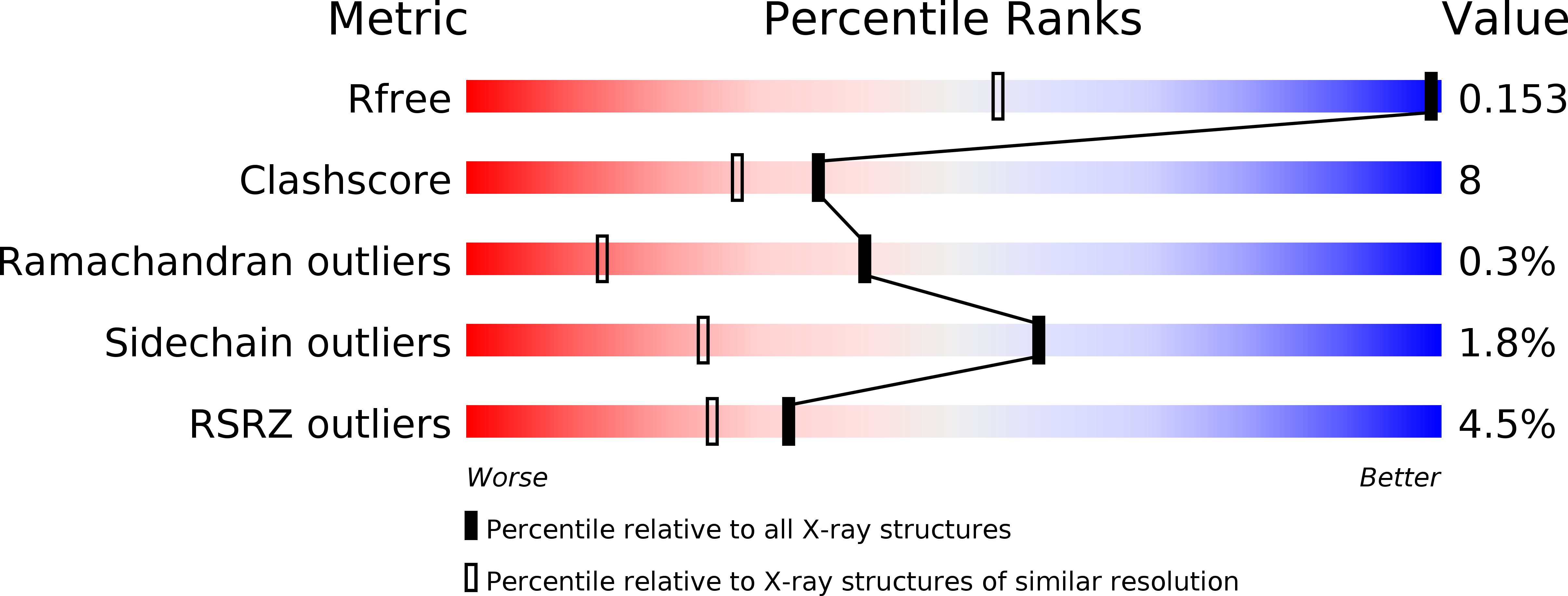
2Z6W image
Deposition Date 2007-08-09
Release Date 2008-04-29
Last Version Date 2023-11-15
Entry Detail
PDB ID:
2Z6W
Title:
Crystal structure of human cyclophilin D in complex with cyclosporin A
Biological Source:
Source Organism:
Method Details:
Experimental Method:
Resolution:
0.96 Å
R-Value Free:
0.15
R-Value Work:
0.12
R-Value Observed:
0.13
Space Group:
P 21 21 21
Macromolecular Entities
Structures with similar UniProt ID
Protein Blast
Polymer Type:polypeptide(L)
Molecule:PEPTIDYL-PROLYL CIS-TRANS ISOMERASE
Gene (Uniprot):PPIF
Mutagens:YES
Chain IDs:A, B
Chain Length:165
Number of Molecules:2
Biological Source:HOMO SAPIENS
Polymer Type:polypeptide(L)
Molecule:CYCLOSPORIN A
Chain IDs:C (auth: M), D (auth: N)
Chain Length:11
Number of Molecules:2
Biological Source:TOLYPOCLADIUM INFLATUM
Ligand Molecules
Peptide-like Molecules
PRD_000142
Primary Citation
Crystal Structure of Human Cyclophilin D in Complex with its Inhibitor, Cyclosporin a at 0.96-A Resolution.
Proteins 70 1635 ? (2008)
PMID: 18076075 DOI: 10.1002/PROT.21855