2Z63 image
Deposition Date 2007-07-22
Release Date 2007-09-18
Last Version Date 2024-11-13
Entry Detail
PDB ID:
2Z63
Keywords:
Title:
Crystal structure of the TV8 hybrid of human TLR4 and hagfish VLRB.61
Biological Source:
Source Organism:
Homo sapiens (Taxon ID: 9606)
Eptatretus burgeri (Taxon ID: 7764)
Host Organism:
Method Details:
Experimental Method:
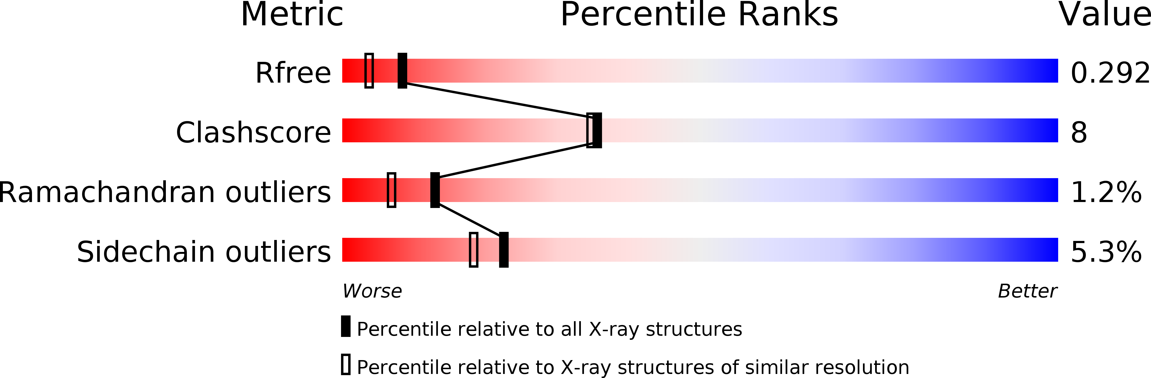
Resolution:
2.00 Å
R-Value Free:
0.29
R-Value Work:
0.24
R-Value Observed:
0.25
Space Group:
P 31
Macromolecular Entities
Polymer Type:polypeptide(L)
Molecule:Toll-like receptor 4, Variable lymphocyte receptor B
Gene (Uniprot):TLR4, VLRB
Chain IDs:A
Chain Length:570
Number of Molecules:1
Biological Source:Homo sapiens, Eptatretus burgeri
Modified Residue
Compound ID Chain ID Parent Comp ID Details 2D Image
ASN A ASN GLYCOSYLATION SITE
Primary Citation
Crystal Structure of the TLR4-MD-2 Complex with Bound Endotoxin Antagonist Eritoran
Cell(Cambridge,Mass.) 130 906 917 (2007)
PMID: 17803912 DOI: 10.1016/j.cell.2007.08.002

Abstact

TLR4 and MD-2 form a heterodimer that recognizes LPS (lipopolysaccharide) from Gram-negative bacteria. Eritoran is an analog of LPS that antagonizes its activity by binding to the TLR4-MD-2 complex. We determined the structure of the full-length ectodomain of the mouse TLR4 and MD-2 complex. We also produced a series of hybrids of human TLR4 and hagfish VLR and determined their structures with and without bound MD-2 and Eritoran. TLR4 is an atypical member of the LRR family and is composed of N-terminal, central, and C-terminal domains. The beta sheet of the central domain shows unusually small radii and large twist angles. MD-2 binds to the concave surface of the N-terminal and central domains. The interaction with Eritoran is mediated by a hydrophobic internal pocket in MD-2. Based on structural analysis and mutagenesis experiments on MD-2 and TLR4, we propose a model of TLR4-MD-2 dimerization induced by LPS.

Legend

Protein

Chemical

Disease

Primary Citation of related structures