2Z5X image
Deposition Date 2007-07-20
Release Date 2008-04-01
Last Version Date 2023-11-01
Entry Detail
PDB ID:
2Z5X
Keywords:
Title:
Crystal Structure of Human Monoamine Oxidase A with Harmine
Biological Source:
Source Organism:
Homo sapiens (Taxon ID: 9606)
Method Details:
Experimental Method:
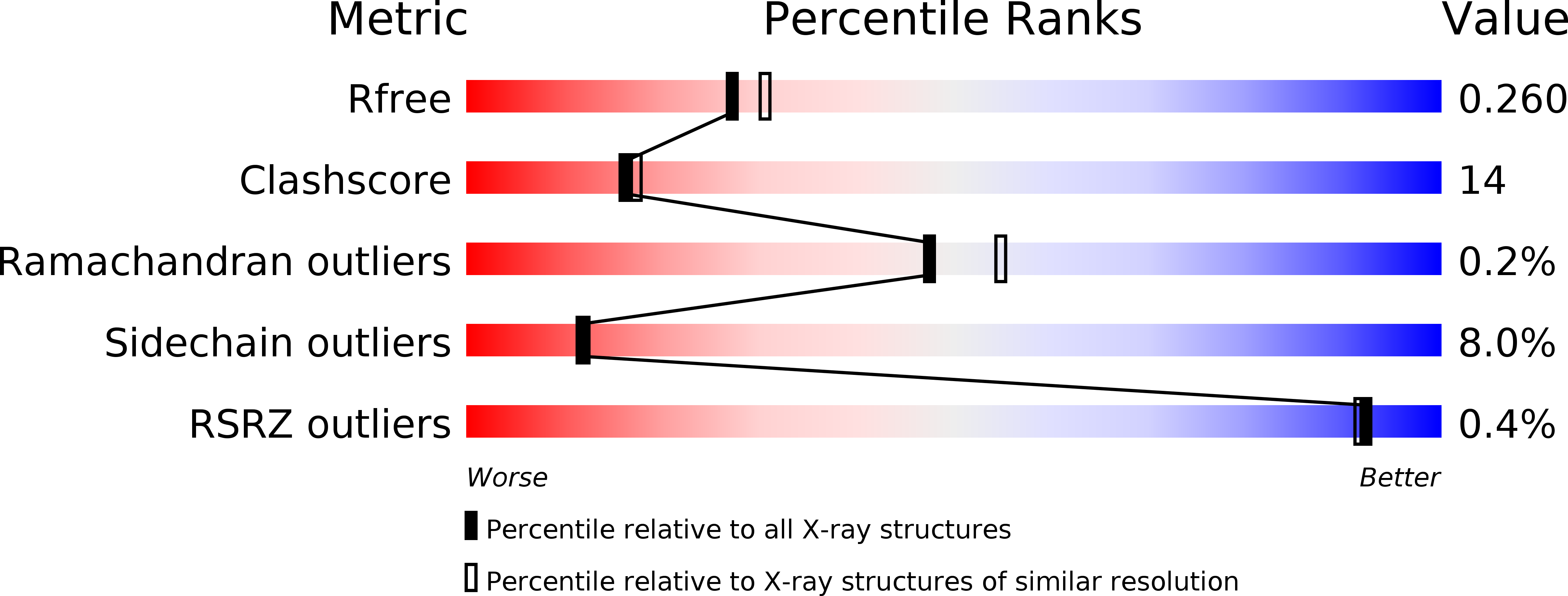
Resolution:
2.20 Å
R-Value Free:
0.25
R-Value Work:
0.20
R-Value Observed:
0.20
Space Group:
C 2 2 2
Macromolecular Entities
Polymer Type:polypeptide(L)
Molecule:Amine oxidase [flavin-containing] A
Gene (Uniprot):MAOA
Chain IDs:A
Chain Length:513
Number of Molecules:1
Biological Source:Homo sapiens
Primary Citation
Structure of human monoamine oxidase A at 2.2-A resolution: The control of opening the entry for substrates/inhibitors
Proc.Natl.Acad.Sci.Usa 105 5739 5744 (2008)
PMID: 18391214 DOI: 10.1073/pnas.0710626105

Abstact

The mitochondrial outer membrane-anchored monoamine oxidase (MAO) is a biochemically important flavoenzyme that catalyzes the deamination of biogenic and xenobiotic amines. Its two subtypes, MAOA and MAOB, are linked to several psychiatric disorders and therefore are interesting targets for drug design. To understand the relationship between structure and function of this enzyme, we extended our previous low-resolution rat MAOA structure to the high-resolution wild-type and G110A mutant human MAOA structures at 2.2 and 2.17 A, respectively. The high-resolution MAOA structures are similar to those of rat MAOA and human MAOB, but different from the known structure of human MAOA [De Colibus L, et al. (2005) Proc Natl Acad Sci USA 102:12684-12689], specifically regarding residues 108-118 and 210-216, which surround the substrate/inhibitor cavity. The results confirm that the inhibitor selectivity of MAOA and MAOB is caused by the structural differences arising from Ile-335 in MAOA vs. Tyr-326 in MAOB. The structures exhibit a C-terminal transmembrane helix with clear electron density, as is also seen in rat MAOA. Mutations on one residue of loop 108-118, G110, which is far from the active center but close to the membrane surface, cause the solubilized enzyme to undergo a dramatic drop in activity, but have less effect when the enzyme is anchored in the membrane. These results suggest that the flexibility of loop 108-118, facilitated by anchoring the enzyme into the membrane, is essential for controlling substrate access to the active site. We report on the observation of the structure-function relationship between a transmembrane helical anchor and an extra-membrane domain.

Legend

Protein

Chemical

Disease

Primary Citation of related structures