Deposition Date
2007-05-25
Release Date
2007-06-19
Last Version Date
2024-07-10
Entry Detail
PDB ID:
2Z2P
Keywords:
Title:
Crystal Structure of catalytically inactive H270A virginiamycin B lyase from Staphylococcus aureus with Quinupristin
Biological Source:
Source Organism:
Staphylococcus aureus (Taxon ID: 1280)
STREPTOMYCES GRAMINOFACIEN (Taxon ID: 68212)
STREPTOMYCES GRAMINOFACIEN (Taxon ID: 68212)
Host Organism:
Method Details:
Experimental Method:
Resolution:
2.80 Å
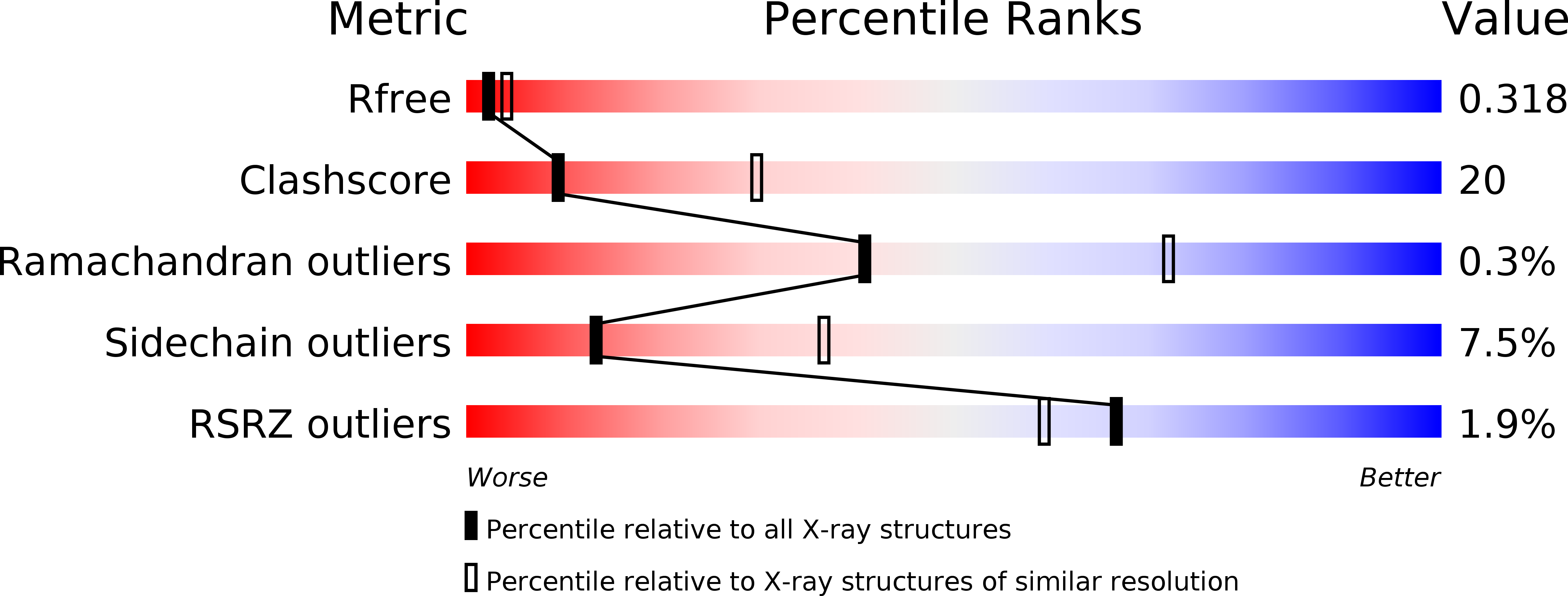
R-Value Free:
0.31
R-Value Work:
0.26
R-Value Observed:
0.26
Space Group:
P 21 21 21