2Z0P image
Entry Detail
PDB ID:
2Z0P
Title:
Crystal structure of PH domain of Bruton's tyrosine kinase
Biological Source:
Source Organism:
Host Organism:
PDB Version:
Deposition Date:
2007-05-07
Release Date:
2008-05-13
Method Details:
Experimental Method:
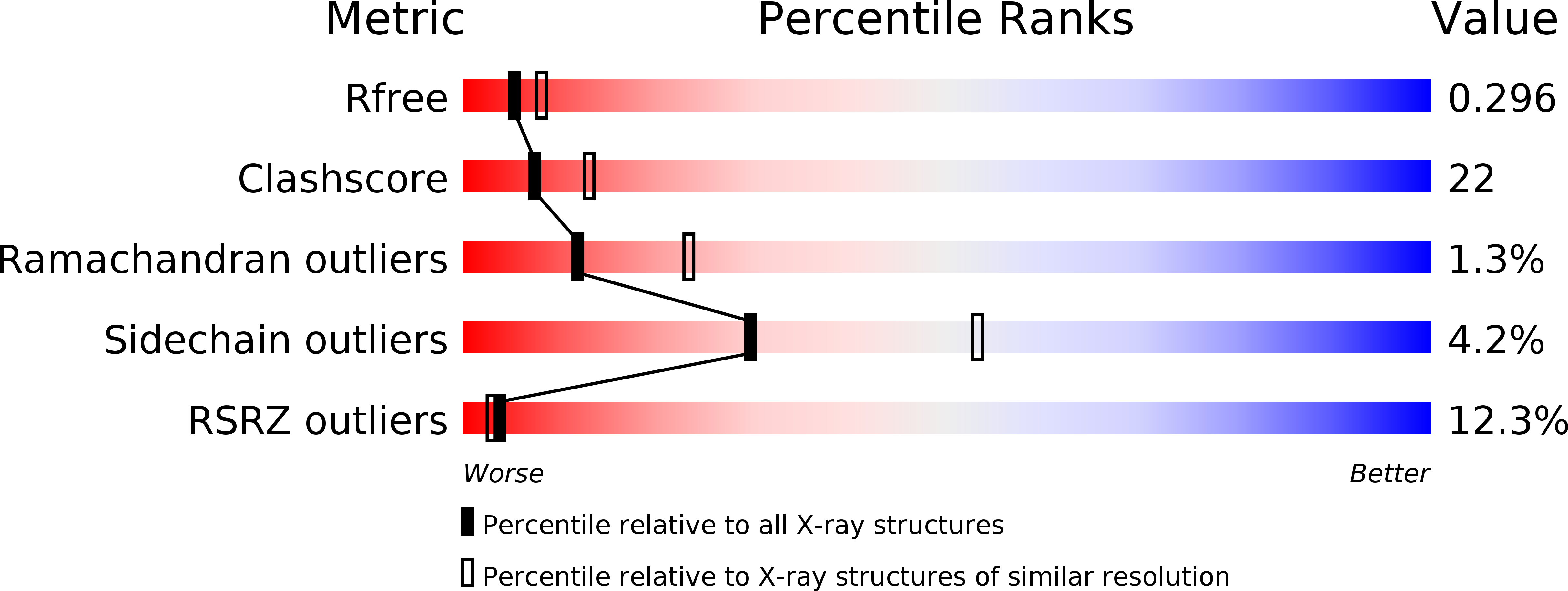
Resolution:
2.58 Å
R-Value Free:
0.3
R-Value Work:
0.24
R-Value Observed:
0.24
Space Group:
P 1 21 1
Macromolecular Entities
Polymer Type:polypeptide(L)
Description:Tyrosine-protein kinase BTK
Chain IDs:A, B, C, D
Chain Length:169
Number of Molecules:4
Biological Source:Homo sapiens
Primary Citation
Crystal structure of the Bruton's tyrosine kinase PH domain with phosphatidylinositol
Biochem.Biophys.Res.Commun. 377 23 28 (2008)
PMID: 18809383 DOI: 10.1016/j.bbrc.2008.09.055

Abstact

Bruton's tyrosine kinase (Btk) of the Tec family possesses a Pleckstrin homology (PH) domain, which is responsible for plasma membrane targeting. In this study, the crystal structure of the Btk PH domain in complex with dibutylyl-phosphatidylinositol-3,4,5-triphosphate was determined. The structure revealed that the Btk PH domain forms a homodimer and that each molecule binds phosphatidylinositol in the binding pocket. The side chain of Lys18 within a Btk-specific insertion in the beta1-beta2 loop is able to form a hydrogen bond with the diacylglycerol moiety of phosphatidylinositol. The other Btk-specific insertion in the beta5-beta6 loop constitutes the dimerization interface. Thus, the modes of phosphatidylinositol recognition and Btk PH domain dimerization are distinct from those of other PH domains.

Legend

Protein

Chemical

Disease

Primary Citation of related structures