Deposition Date
2007-05-07
Release Date
2007-05-22
Last Version Date
2023-11-01
Entry Detail
PDB ID:
2Z0D
Keywords:
Title:
The crystal structure of human Atg4B- LC3(1-120) complex
Biological Source:
Source Organism(s):
Homo sapiens (Taxon ID: 9606)
Rattus norvegicus (Taxon ID: 10116)
Rattus norvegicus (Taxon ID: 10116)
Expression System(s):
Method Details:
Experimental Method:
Resolution:
1.90 Å
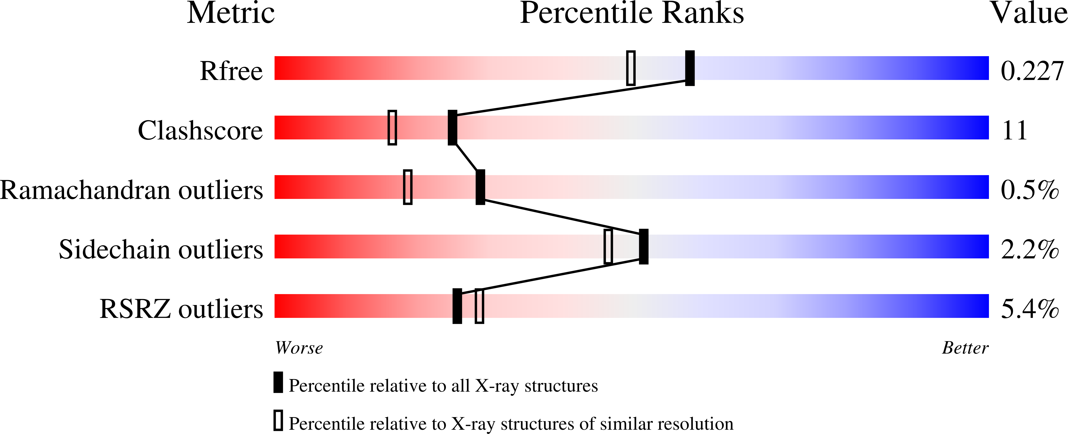
R-Value Free:
0.22
R-Value Work:
0.19
R-Value Observed:
0.19
Space Group:
P 21 21 21