Deposition Date
2007-05-06
Release Date
2007-11-06
Last Version Date
2023-10-25
Entry Detail
PDB ID:
2Z01
Keywords:
Title:
Crystal structure of phosphoribosylaminoimidazole synthetase from Geobacillus kaustophilus
Biological Source:
Source Organism(s):
Geobacillus kaustophilus (Taxon ID: 1462)
Expression System(s):
Method Details:
Experimental Method:
Resolution:
2.20 Å
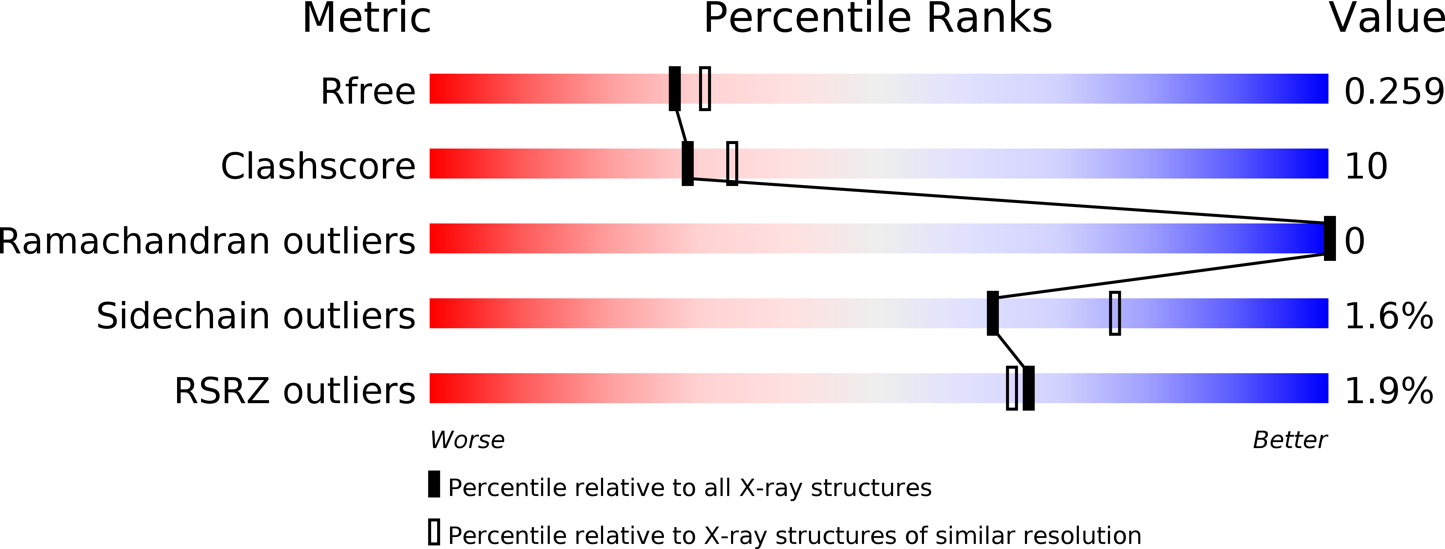
R-Value Free:
0.25
R-Value Work:
0.22
R-Value Observed:
0.22
Space Group:
P 21 21 2