Deposition Date
2007-04-13
Release Date
2008-04-01
Last Version Date
2023-11-08
Entry Detail
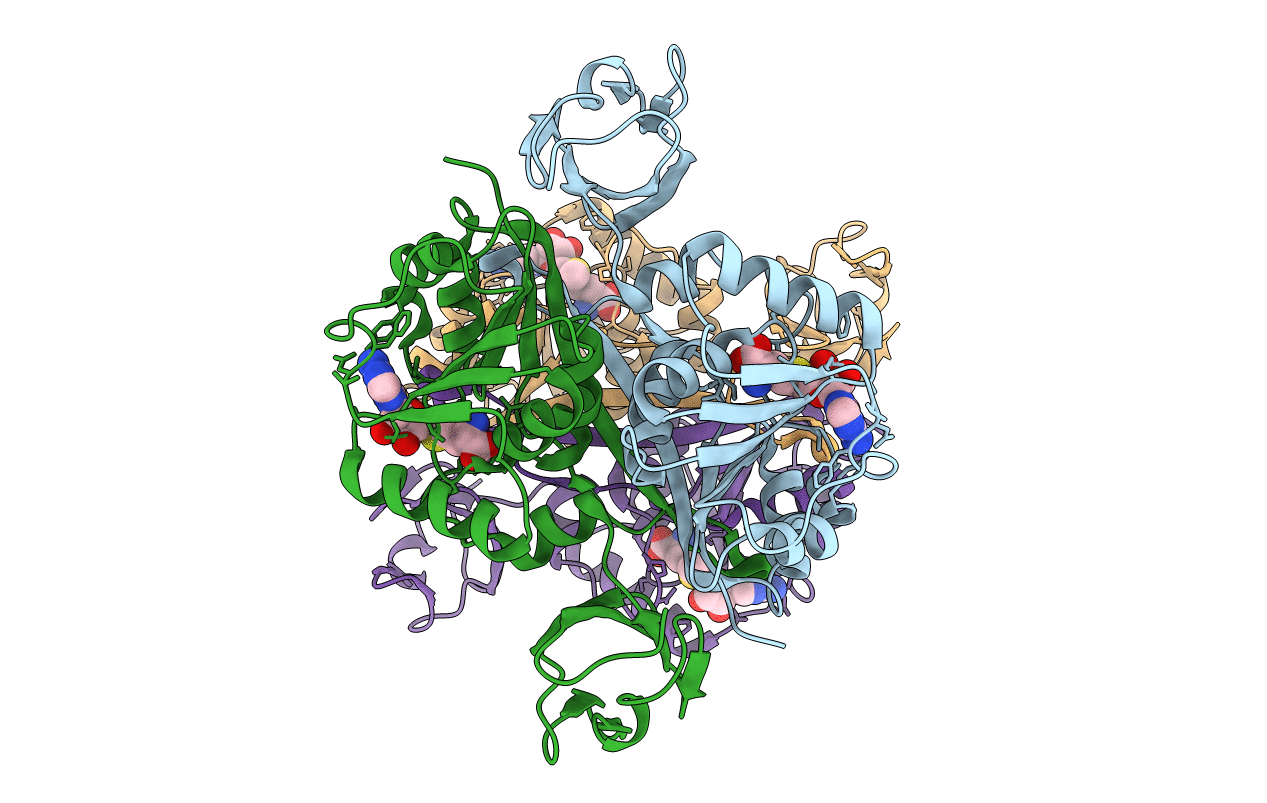
PDB ID:
2YVL
Keywords:
Title:
Crystal structure of tRNA (m1A58) methyltransferase TrmI from Aquifex aeolicus
Biological Source:
Source Organism(s):
Aquifex aeolicus (Taxon ID: 224324)
Expression System(s):
Method Details:
Experimental Method:
Resolution:
2.20 Å
R-Value Free:
0.22
R-Value Work:
0.19
R-Value Observed:
0.19
Space Group:
P 21 21 21