2YV9 image
Deposition Date 2007-04-10
Release Date 2008-02-19
Last Version Date 2023-10-25
Entry Detail
PDB ID:
2YV9
Keywords:
Title:
Crystal structure of the CLIC homologue EXC-4 from c. elegans
Biological Source:
Source Organism:
Host Organism:
Method Details:
Experimental Method:
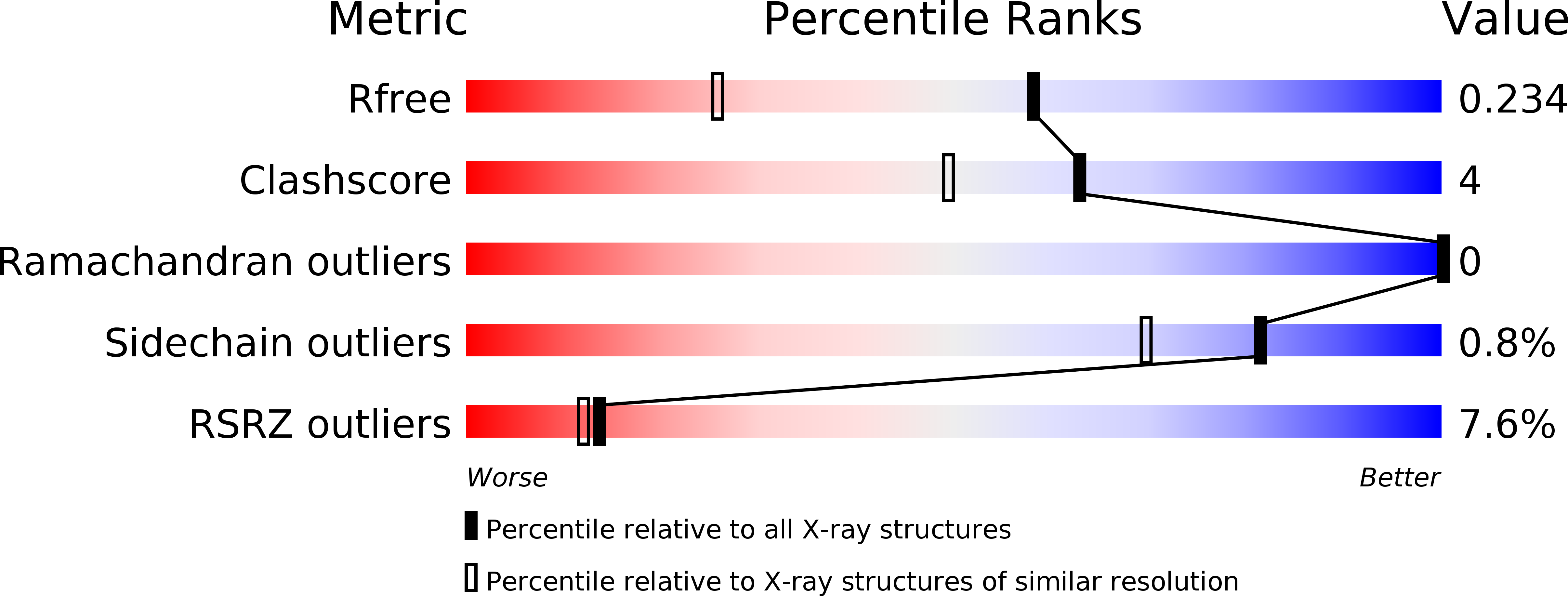
Resolution:
1.60 Å
R-Value Free:
0.23
R-Value Work:
0.18
R-Value Observed:
0.19
Space Group:
P 1 21 1
Macromolecular Entities
Polymer Type:polypeptide(L)
Molecule:Chloride intracellular channel exc-4
Gene (Uniprot):exc-4
Chain IDs:A, B
Chain Length:291
Number of Molecules:2
Biological Source:Caenorhabditis elegans
Ligand Molecules
Primary Citation
Comparison of vertebrate and invertebrate CLIC proteins: The crystal structures of Caenorhabditis elegans EXC-4 and Drosophila melanogaster DmCLIC
Proteins 71 364 378 (2007)
PMID: 17985355 DOI: 10.1002/prot.21704

Abstact

The crystal structures of two CLIC family members DmCLIC and EXC-4 from the invertebrates Drosophila melanogaster and Caenorhabditis elegans, respectively, have been determined. The proteins adopt a glutathione S-transferase (GST) fold. The structures are highly homologous to each other and more closely related to the known structures of the human CLIC1 and CLIC4 than to GSTs. The invertebrate CLICs show several unique features including an elongated C-terminal extension and a divalent metal binding site. The latter appears to alter the ancestral glutathione binding site, and thus, the invertebrate CLICs are unlikely to bind glutathione in the same manner as the GST proteins. Purified recombinant DmCLIC and EXC-4 both bind to lipid bilayers and can form ion channels in artificial lipid bilayers, albeit at low pH. EXC-4 differs from other CLIC proteins in that the conserved redox-active cysteine at the N-terminus of helix 1 is replaced by an aspartic acid residue. Other key distinguishing features of EXC-4 include the fact that it binds to artificial bilayers at neutral pH and this binding is not sensitive to oxidation. These differences with other CLIC family members are likely to be due to the substitution of the conserved cysteine by aspartic acid.

Legend

Protein

Chemical

Disease

Primary Citation of related structures