2YT1 image
Entry Detail
PDB ID:
2YT1
Keywords:
Title:
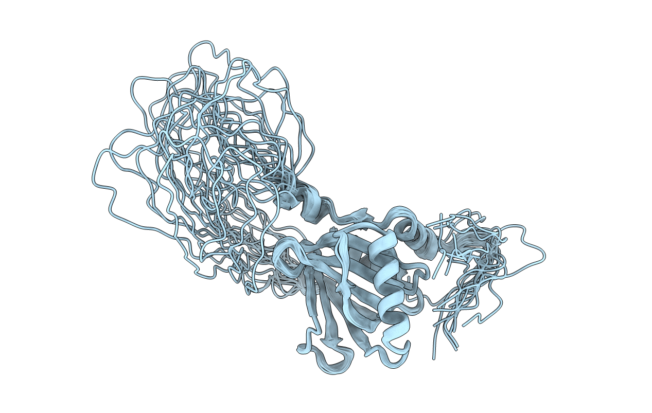
Solution structure of the chimera of the C-terminal tail peptide of APP and the C-terminal PID domain of Fe65L
Biological Source:
Source Organism:
PDB Version:
Deposition Date:
2007-04-05
Release Date:
2008-04-08
Method Details:
Experimental Method:
Conformers Calculated:
100
Conformers Submitted:
20
Selection Criteria:
structures with the least restraint violations, target function
Macromolecular Entities
Polymer Type:polypeptide(L)
Description:Amyloid beta A4 protein and Amyloid beta A4 precursor protein-binding family B member 2
Chain IDs:A
Chain Length:185
Number of Molecules:1
Biological Source:Mus musculus
Ligand Molecules
Primary Citation
Structure of the C-terminal phosphotyrosine interaction domain of Fe65L1 complexed with the cytoplasmic tail of amyloid precursor protein reveals a novel peptide binding mode
J.Biol.Chem. 283 27165 27178 (2008)
PMID: 18650440 DOI: 10.1074/jbc.M803892200

Abstact

Fe65L1, a member of the Fe65 family, is an adaptor protein that interacts with the cytoplasmic domain of Alzheimer amyloid precursor protein (APP) through its C-terminal phosphotyrosine interaction/phosphotyrosine binding (PID/PTB) domain. In the present study, the solution structures of the C-terminal PID domain of mouse Fe65L1, alone and in complex with a 32-mer peptide (DAAVTPEERHLSKMQQNGYENPTYKFFEQMQN) derived from the cytoplasmic domain of APP, were determined using NMR spectroscopy. The C-terminal PID domain of Fe65L1 alone exhibits a canonical PID/PTB fold, whereas the complex structure reveals a novel mode of peptide binding. In the complex structure, the NPTY motif forms a type-I beta-turn, and the residues immediately N-terminal to the NPTY motif form an antiparallel beta-sheet with the beta5 strand of the PID domain, the binding mode typically observed in the PID/PTB.peptide complex. On the other hand, the N-terminal region of the peptide forms a 2.5-turn alpha-helix and interacts extensively with the C-terminal alpha-helix and the peripheral regions of the PID domain, representing a novel mode of peptide binding that has not been reported previously for the PID/PTB.peptide complex. The indispensability of the N-terminal region of the peptide for the high affinity of the PID-peptide interaction is consistent with NMR titration and isothermal calorimetry data. The extensive binding features of the PID domain of Fe65L1 with the cytoplasmic domain of APP provide a framework for further understanding of the function, trafficking, and processing of APP modulated by adapter proteins.

Legend

Protein

Chemical

Disease

Primary Citation of related structures