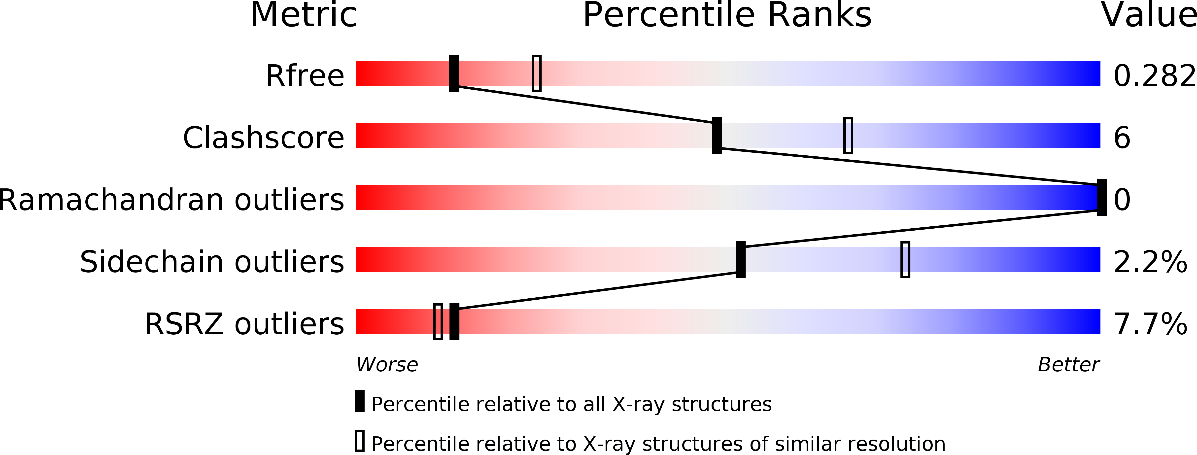
2YQ2 image
Deposition Date 2012-11-04
Release Date 2013-01-16
Last Version Date 2024-10-23
Entry Detail
PDB ID:
2YQ2
Keywords:
Title:
Structure of BVDV1 envelope glycoprotein E2, pH8
Biological Source:
Source Organism:
Host Organism:
Method Details:
Experimental Method:
Resolution:
2.58 Å
R-Value Free:
0.25
R-Value Work:
0.23
R-Value Observed:
0.24
Space Group:
C 1 2 1
Macromolecular Entities
Polymer Type:polypeptide(L)
Molecule:BVDV1 E2
Mutations:YES
Chain IDs:A, B
Chain Length:337
Number of Molecules:2
Biological Source:BOVINE VIRAL DIARRHEA VIRUS 1
Modified Residue
Compound ID Chain ID Parent Comp ID Details 2D Image
ASN A ASN GLYCOSYLATION SITE
Ligand Molecules
Primary Citation
Structure of a Pestivirus Envelope Glycoprotein E2 Clarifies its Role in Cell Entry.
Cell Rep. 3 30 ? (2013)
PMID: 23273918 DOI: 10.1016/J.CELREP.2012.12.001

Abstact

Enveloped viruses have developed various adroit mechanisms to invade their host cells. This process requires one or more viral envelope glycoprotein to achieve cell attachment and membrane fusion. Members of the Flaviviridae such as flaviviruses possess only one envelope glycoprotein, E, whereas pestiviruses and hepacivirus encode two glycoproteins, E1 and E2. Although E2 is involved in cell attachment, it has been unclear which protein is responsible for membrane fusion. We report the crystal structures of the homodimeric glycoprotein E2 from the pestivirus bovine viral diarrhea virus 1 (BVDV1) at both neutral and low pH. Unexpectedly, BVDV1 E2 does not have a class II fusion protein fold, and at low pH the N-terminal domain is disordered, similarly to the intermediate postfusion state of E2 from sindbis virus, an alphavirus. Our results suggest that the pestivirus and possibly the hepacivirus fusion machinery are unlike any previously observed.

Legend

Protein

Chemical

Disease

Primary Citation of related structures