Deposition Date
2012-11-02
Release Date
2013-11-13
Last Version Date
2024-05-08
Entry Detail
PDB ID:
2YQ1
Keywords:
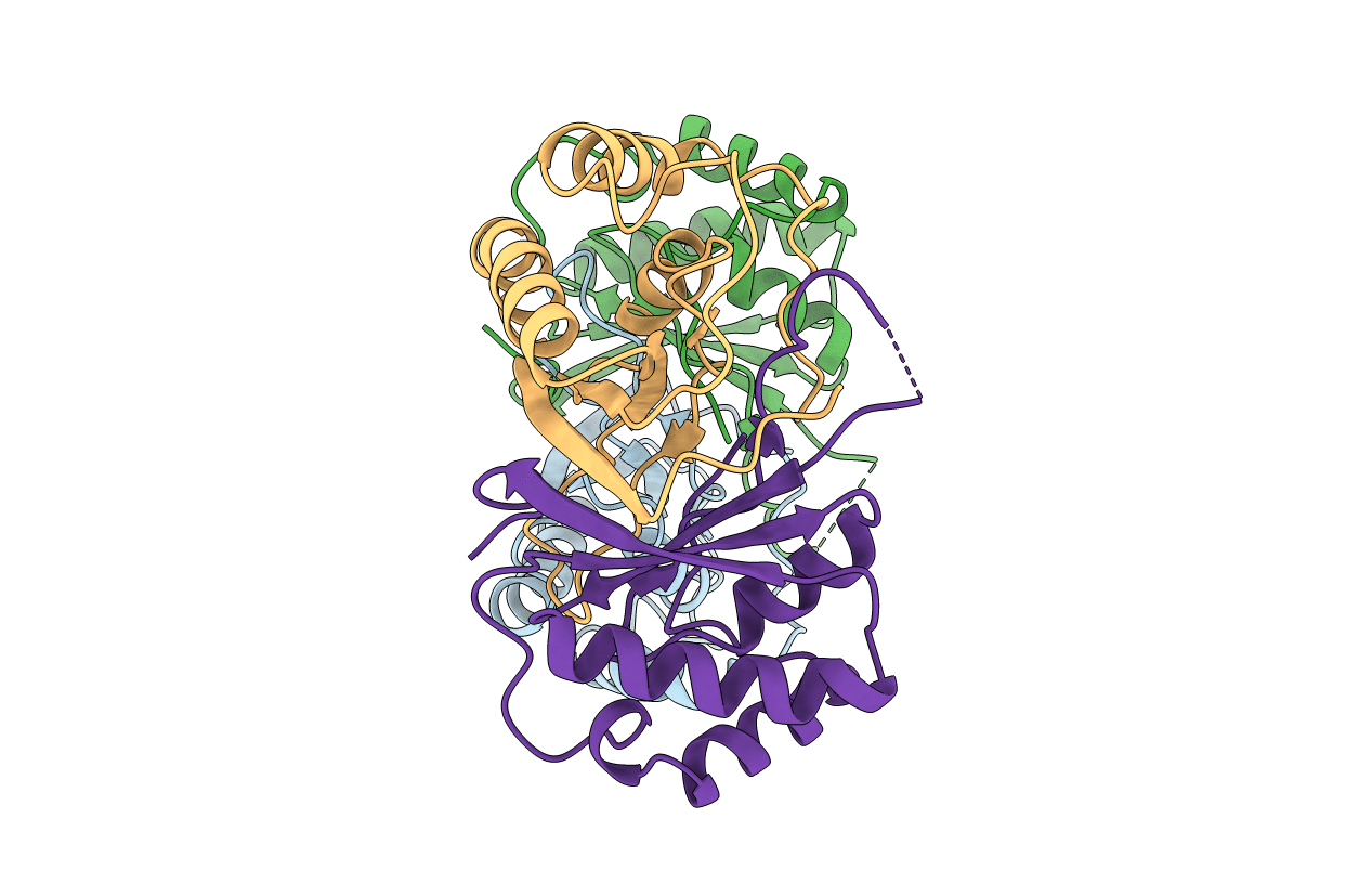
Title:
MHV-68 LANA (ORF73) C-terminal domain: triclinic crystal form
Biological Source:
Source Organism(s):
MURID HERPESVIRUS 4 (Taxon ID: 33708)
Expression System(s):
Method Details:
Experimental Method:
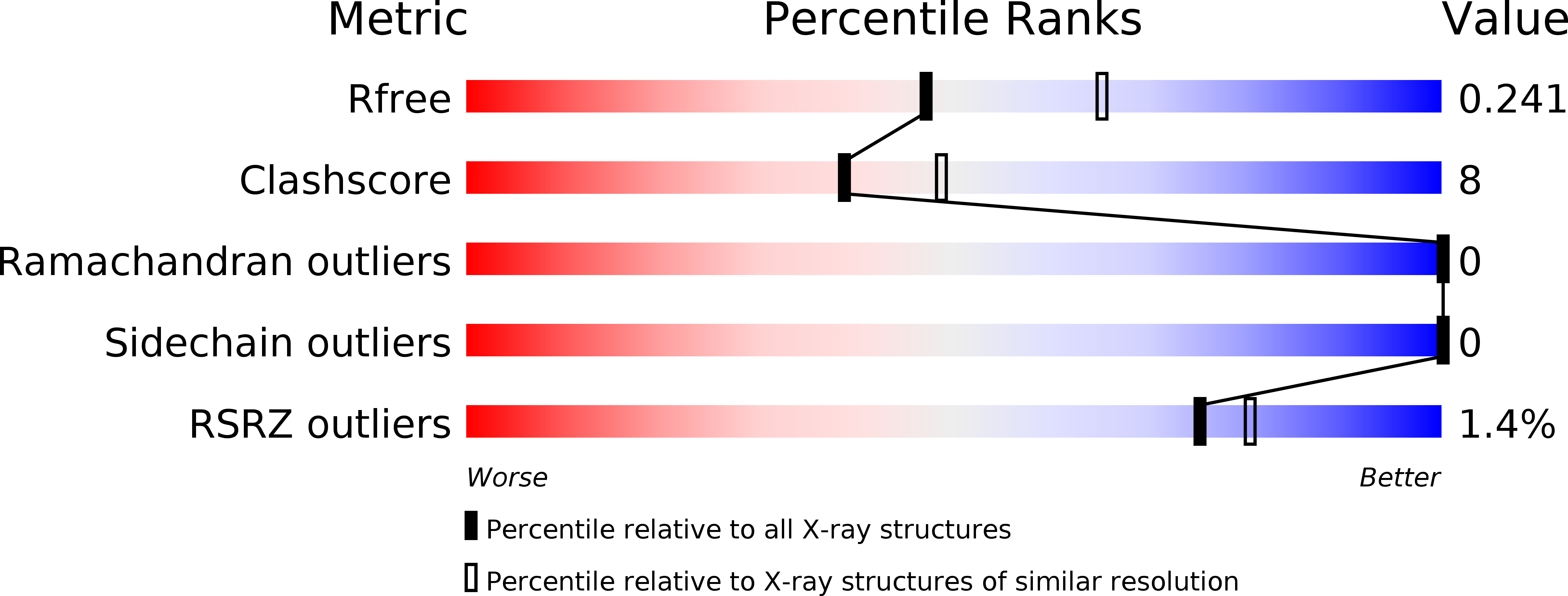
Resolution:
2.30 Å
R-Value Free:
0.24
R-Value Work:
0.20
R-Value Observed:
0.20
Space Group:
P 1