
Deposition Date
2012-10-29
Release Date
2013-04-10
Last Version Date
2024-05-08
Entry Detail
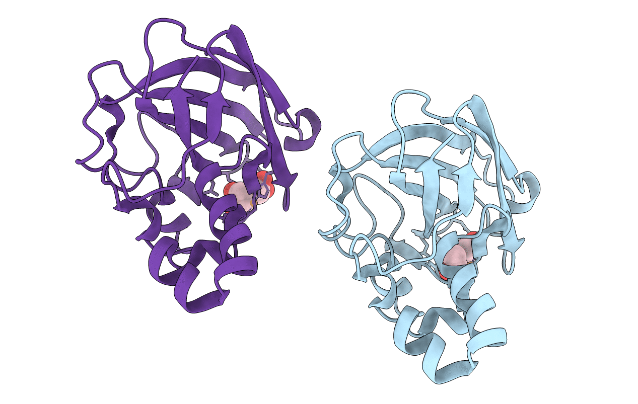
PDB ID:
2YOY
Keywords:
Title:
Bacillus amyloliquefaciens CBM33 in complex with Cu(I) reduced using ascorbate
Biological Source:
Source Organism(s):
BACILLUS AMYLOLIQUEFACIENS (Taxon ID: 1390)
Expression System(s):
Method Details:
Experimental Method:
Resolution:
1.70 Å
R-Value Free:
0.24
R-Value Work:
0.20
R-Value Observed:
0.21
Space Group:
P 21 21 21
