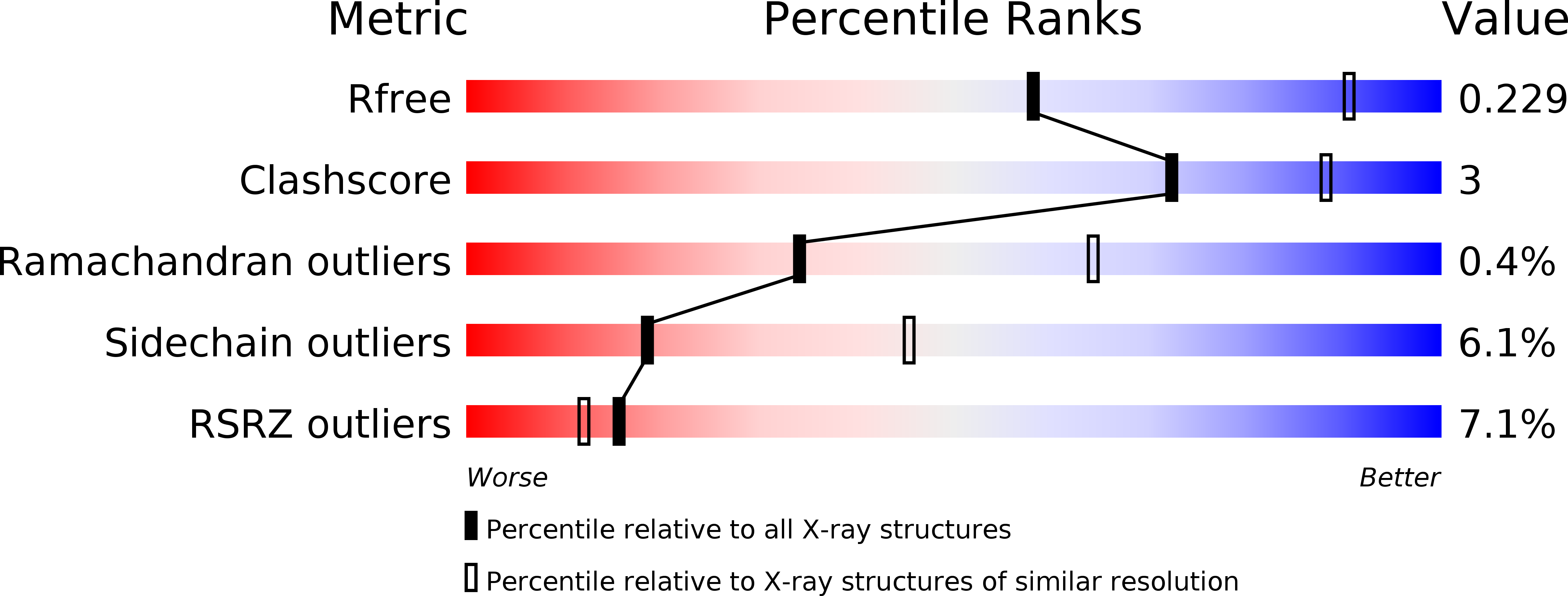
2YOC image
Entry Detail
PDB ID:
2YOC
Keywords:
Title:
Crystal structure of PulA from Klebsiella oxytoca
Biological Source:
Source Organism:
Host Organism:
PDB Version:
Deposition Date:
2012-10-23
Release Date:
2013-11-06
Method Details:
Experimental Method:
Resolution:
2.88 Å
R-Value Free:
0.21
R-Value Work:
0.17
R-Value Observed:
0.17
Space Group:
P 63 2 2
Macromolecular Entities
Polymer Type:polypeptide(L)
Description:PULLULANASE
Chain IDs:A, B
Chain Length:1078
Number of Molecules:2
Biological Source:KLEBSIELLA OXYTOCA
Modified Residue
Compound ID Chain ID Parent Comp ID Details 2D Image
MSE A MET SELENOMETHIONINE
Primary Citation
Structural Basis of Pullulanase Membrane Binding and Secretion Revealed by X-Ray Crystallography, Molecular Dynamics and Biochemical Analysis
Structure 24 92 ? (2016)
PMID: 26688215 DOI: 10.1016/J.STR.2015.10.023

Abstact

The Klebsiella lipoprotein pullulanase (PulA) is exported to the periplasm, triacylated, and anchored via lipids in the inner membrane (IM) prior to its transport to the bacterial surface through a type II secretion system (T2SS). X-Ray crystallography and atomistic molecular dynamics (MD) simulations of PulA in a 1-palmitoyl-2-oleoyl-sn-glycero-3-phosphoethanolamine (POPE) model membrane provided an unprecedented molecular view of an N-terminal unstructured tether and the IM lipoprotein retention signal, and revealed novel interactions with the IM via N-terminal immunoglobulin-like domains in PulA. An efficiently secreted nonacylated variant (PulANA) showed similar peripheral membrane association during MD simulations, consistent with the binding of purified PulANA to liposomes. Remarkably, combined X-ray, MD, and functional studies identified a novel subdomain, Ins, inserted in the α-amylase domain, which is required for PulA secretion. Available data support a model in which PulA binding to the IM promotes interactions with the T2SS, possibly via the Ins subdomain.

Legend

Protein

Chemical

Disease

Primary Citation of related structures