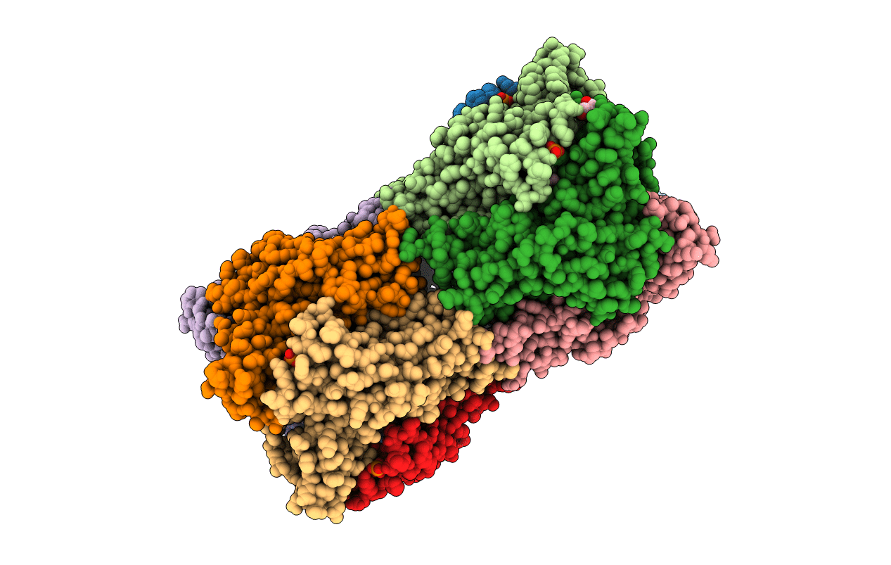
2YMD image
Entry Detail
PDB ID:
2YMD
Keywords:
Title:
Crystal structure of a mutant binding protein (5HTBP-AChBP) in complex with serotonin (5-hydroxytryptamine)
Biological Source:
Source Organism:
Host Organism:
PDB Version:
Deposition Date:
2012-10-09
Release Date:
2012-12-12
Method Details:
Experimental Method:
Resolution:
1.96 Å
R-Value Free:
0.20
R-Value Work:
0.15
R-Value Observed:
0.15
Space Group:
C 1 2 1
Macromolecular Entities
Polymer Type:polypeptide(L)
Description:SOLUBLE ACETYLCHOLINE RECEPTOR
Mutations:YES
Chain IDs:A, B, C, D, E, F, G, H, I, J
Chain Length:212
Number of Molecules:10
Biological Source:APLYSIA CALIFORNICA
Primary Citation
Structural Basis of Ligand Recognition in 5-Ht(3) Receptors.
Embo Rep. 14 49 ? (2013)
PMID: 23196367 DOI: 10.1038/EMBOR.2012.189

Abstact

The 5-HT(3) receptor is a pentameric serotonin-gated ion channel, which mediates rapid excitatory neurotransmission and is the target of a therapeutically important class of anti-emetic drugs, such as granisetron. We report crystal structures of a binding protein engineered to recognize the agonist serotonin and the antagonist granisetron with affinities comparable to the 5-HT(3) receptor. In the serotonin-bound structure, we observe hydrophilic interactions with loop E-binding site residues, which might enable transitions to channel opening. In the granisetron-bound structure, we observe a critical cation-π interaction between the indazole moiety of the ligand and a cationic centre in loop D, which is uniquely present in the 5-HT(3) receptor. We use a series of chemically tuned granisetron analogues to demonstrate the energetic contribution of this electrostatic interaction to high-affinity ligand binding in the human 5-HT(3) receptor. Our study offers the first structural perspective on recognition of serotonin and antagonism by anti-emetics in the 5-HT(3) receptor.

Legend

Protein

Chemical

Disease