2YJN image
Deposition Date 2011-05-20
Release Date 2011-11-09
Last Version Date 2024-05-08
Entry Detail
PDB ID:
2YJN
Keywords:
Title:
Structure of the glycosyltransferase EryCIII from the erythromycin biosynthetic pathway, in complex with its activating partner, EryCII
Biological Source:
Source Organism:
Host Organism:
Method Details:
Experimental Method:
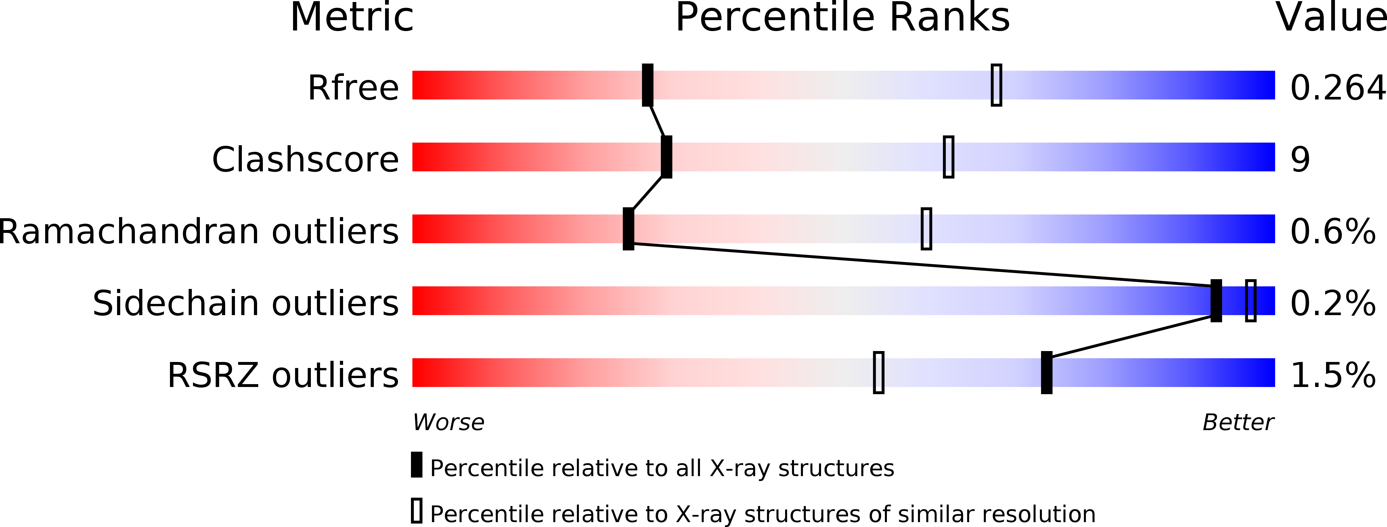
Resolution:
3.09 Å
R-Value Free:
0.26
R-Value Work:
0.21
R-Value Observed:
0.21
Space Group:
P 2 3
Macromolecular Entities
Polymer Type:polypeptide(L)
Molecule:GLYCOSYLTRANSFERASE
Chain IDs:A
Chain Length:441
Number of Molecules:1
Biological Source:SACCHAROPOLYSPORA ERYTHRAEA
Polymer Type:polypeptide(L)
Molecule:DTDP-4-KETO-6-DEOXY-HEXOSE 3,4-ISOMERASE
Chain IDs:B
Chain Length:381
Number of Molecules:1
Biological Source:SACCHAROPOLYSPORA ERYTHRAEA
Ligand Molecules
Primary Citation
Structure of the Glycosyltransferase Eryciii in Complex with its Activating P450 Homologue Erycii.
J.Mol.Biol. 415 92 ? (2012)
PMID: 22056329 DOI: 10.1016/J.JMB.2011.10.036

Abstact

In the biosynthesis of the clinically important antibiotic erythromycin D, the glycosyltransferase (GT) EryCIII, in concert with its partner EryCII, attaches a nucleotide-activated sugar to the macrolide scaffold with high specificity. To understand the role of EryCII, we have determined the crystal structure of the EryCIII·EryCII complex at 3.1 Å resolution. The structure reveals a heterotetramer with a distinctive, elongated quaternary organization. The EryCIII subunits form an extensive self-complementary dimer interface at the center of the complex, and the EryCII subunits lie on the periphery. EryCII binds in the vicinity of the putative macrolide binding site of EryCIII but does not make direct interactions with this site. Our biophysical and enzymatic data support a model in which EryCII stabilizes EryCIII and also functions as an allosteric activator of the GT.

Legend

Protein

Chemical

Disease

Primary Citation of related structures