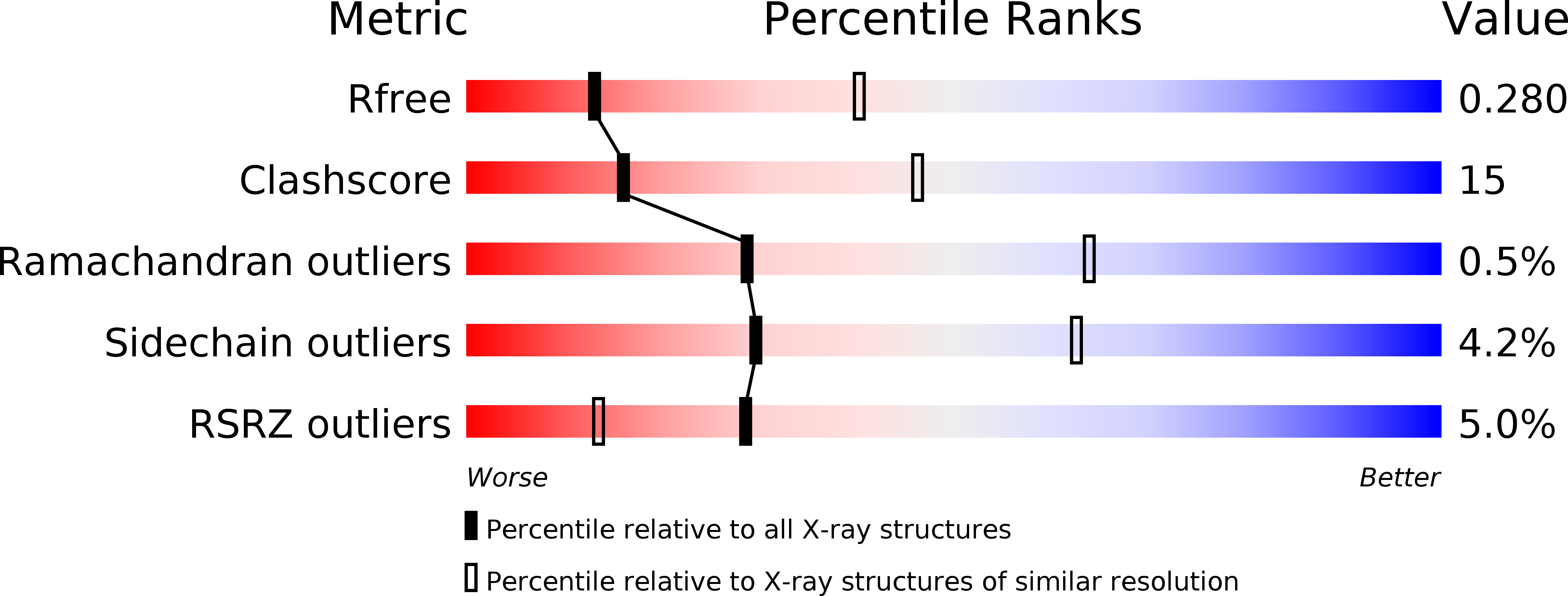
2YJE image
Deposition Date 2011-05-19
Release Date 2011-07-06
Last Version Date 2023-12-20
Entry Detail
PDB ID:
2YJE
Keywords:
Title:
Oligomeric assembly of actin bound to MRTF-A
Biological Source:
Source Organism:
Host Organism:
Method Details:
Experimental Method:
Resolution:
3.10 Å
R-Value Free:
0.27
R-Value Work:
0.23
R-Value Observed:
0.23
Space Group:
P 43 21 2
Macromolecular Entities
Polymer Type:polypeptide(L)
Molecule:ACTIN, ALPHA SKELETAL MUSCLE
Gene (Uniprot):ACTA1
Chain IDs:A, B, C
Chain Length:377
Number of Molecules:3
Biological Source:ORYCTOLAGUS CUNICULUS
Polymer Type:polypeptide(L)
Molecule:MKL/MYOCARDIN-LIKE PROTEIN 1
Gene (Uniprot):Mrtfa
Chain IDs:D (auth: M)
Chain Length:137
Number of Molecules:1
Biological Source:MUS MUSCULUS
Primary Citation
Structure of a pentavalent G-actin*MRTF-A complex reveals how G-actin controls nucleocytoplasmic shuttling of a transcriptional coactivator.
Sci Signal 4 ra40 ra40 (2011)
PMID: 21673315 DOI: 10.1126/scisignal.2001750

Abstact

Subcellular localization of the actin-binding transcriptional coactivator MRTF-A is controlled by its interaction with monomeric actin (G-actin). Signal-induced decreases in G-actin concentration reduce MRTF-A nuclear export, leading to its nuclear accumulation, whereas artificial increases in G-actin concentration in resting cells block MRTF-A nuclear import, retaining it in the cytoplasm. This regulation is dependent on three actin-binding RPEL motifs in the regulatory domain of MRTF-A. We describe the structures of pentavalent and trivalent G-actin•RPEL domain complexes. In the pentavalent complex, each RPEL motif and the two intervening spacer sequences bound an actin monomer, forming a compact assembly. In contrast, the trivalent complex lacked the C-terminal spacer- and RPEL-actins, both of which bound only weakly in the pentavalent complex. Cytoplasmic localization of MRTF-A in unstimulated fibroblasts also required binding of G-actin to the spacer sequences. The bipartite MRTF-A nuclear localization sequence was buried in the pentameric assembly, explaining how increases in G-actin concentration prevent nuclear import of MRTF-A. Analyses of the pentavalent and trivalent complexes show how actin loads onto the RPEL domain and reveal a molecular mechanism by which actin can control the activity of one of its binding partners.

Legend

Protein

Chemical

Disease