2YIU image
Deposition Date 2011-05-16
Release Date 2011-10-26
Last Version Date 2024-10-23
Entry Detail
PDB ID:
2YIU
Keywords:
Title:
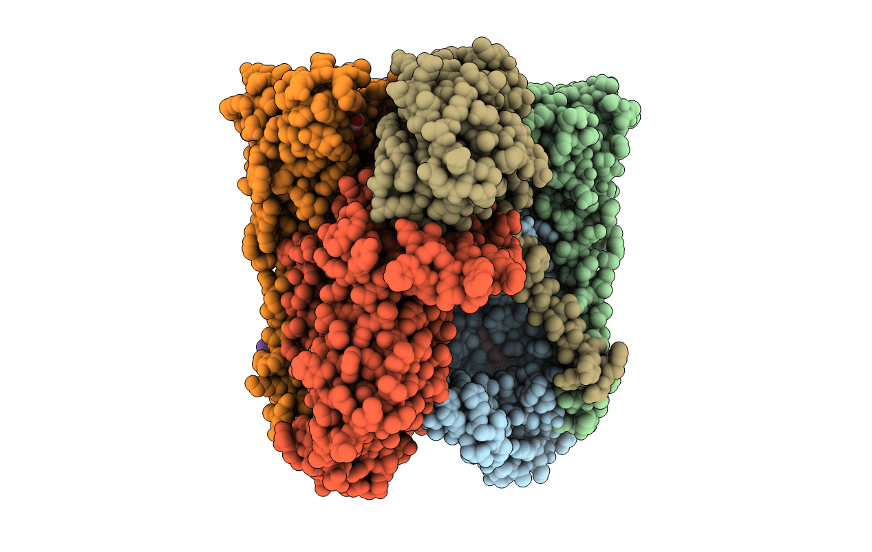
X-ray structure of the dimeric cytochrome BC1 complex from the soil bacterium paracoccus denitrificans at 2.7 angstrom resolution
Biological Source:
Source Organism:
Method Details:
Experimental Method:
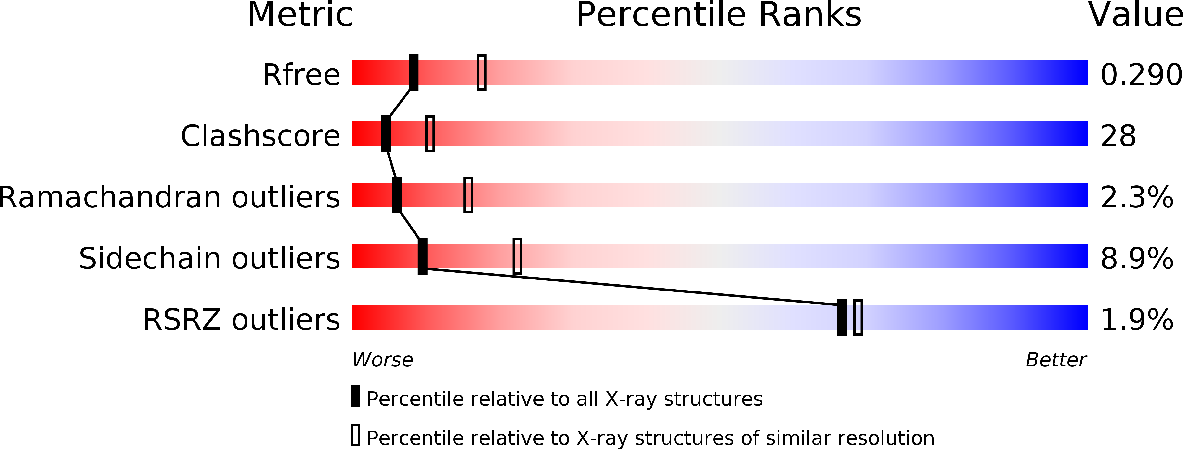
Resolution:
2.70 Å
R-Value Free:
0.29
R-Value Work:
0.23
R-Value Observed:
0.24
Space Group:
P 1 21 1
Macromolecular Entities
Structures with similar UniProt ID
Protein Blast
Polymer Type:polypeptide(L)
Molecule:CYTOCHROME B
Gene (Uniprot):petB
Chain IDs:A, D
Chain Length:450
Number of Molecules:2
Biological Source:PARACOCCUS DENITRIFICANS
Structures with similar UniProt ID
Protein Blast
Polymer Type:polypeptide(L)
Molecule:CYTOCHROME C1, HEME PROTEIN
Gene (Uniprot):petC
Chain IDs:B, E
Chain Length:263
Number of Molecules:2
Biological Source:PARACOCCUS DENITRIFICANS
Structures with similar UniProt ID
Protein Blast
Polymer Type:polypeptide(L)
Molecule:UBIQUINOL-CYTOCHROME C REDUCTASE IRON-SULFUR SUBUNIT
Gene (Uniprot):petA
Chain IDs:C, F
Chain Length:190
Number of Molecules:2
Biological Source:PARACOCCUS DENITRIFICANS
Primary Citation
X-Ray Structure of the Dimeric Cytochrome Bc(1) Complex from the Soil Bacterium Paracoccus Denitrificans at 2.7-A Resolution.
Biochim.Biophys.Acta 1807 1606 ? (2011)
PMID: 21996020 DOI: 10.1016/J.BBABIO.2011.09.017

Abstact

The respiratory cytochrome bc(1) complex is a fundamental enzyme in biological energy conversion. It couples electron transfer from ubiquinol to cytochrome c with generation of proton motive force which fuels ATP synthesis. The complex from the α-proteobacterium Paracoccus denitrificans, a model for the medically relevant mitochondrial complexes, lacked structural characterization. We show by LILBID mass spectrometry that truncation of the organism-specific, acidic N-terminus of cytochrome c(1) changes the oligomerization state of the enzyme to a dimer. The fully functional complex was crystallized and the X-ray structure determined at 2.7-Å resolution. It has high structural homology to mitochondrial complexes and to the Rhodobacter sphaeroides complex especially for subunits cytochrome b and ISP. Species-specific binding of the inhibitor stigmatellin is noteworthy. Interestingly, cytochrome c(1) shows structural differences to the mitochondrial and even between the two Rhodobacteraceae complexes. The structural diversity in the cytochrome c(1) surface facing the ISP domain indicates low structural constraints on that surface for formation of a productive electron transfer complex. A similar position of the acidic N-terminal domains of cytochrome c(1) and yeast subunit QCR6p is suggested in support of a similar function. A model of the electron transfer complex with membrane-anchored cytochrome c(552), the natural substrate, shows that it can adopt the same orientation as the soluble substrate in the yeast complex. The full structural integrity of the P. denitrificans variant underpins previous mechanistic studies on intermonomer electron transfer and paves the way for using this model system to address open questions of structure/function relationships and inhibitor binding.

Legend

Protein

Chemical

Disease

Primary Citation of related structures
Feedback Form
Name
Email
Institute
Feedback