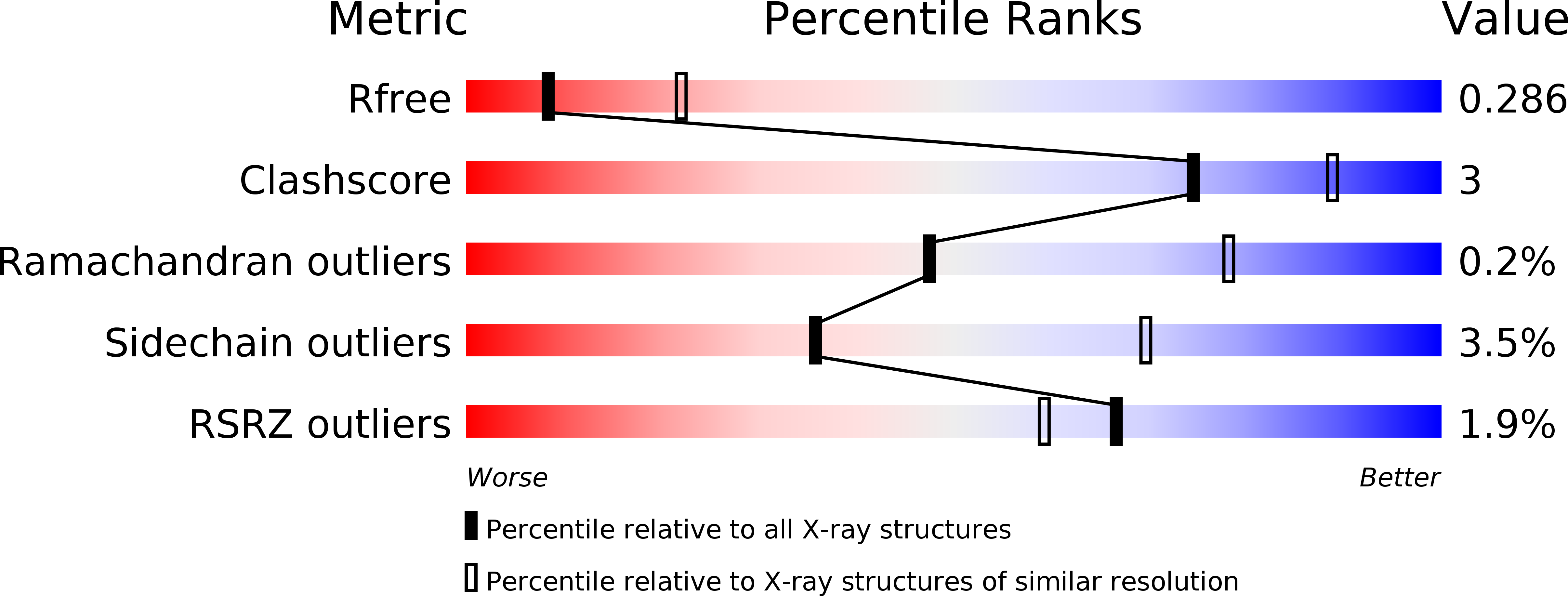
2YGW image
Deposition Date 2011-04-21
Release Date 2012-02-15
Last Version Date 2024-10-16
Entry Detail
PDB ID:
2YGW
Keywords:
Title:
Crystal structure of human MCD
Biological Source:
Source Organism:
HOMO SAPIENS (Taxon ID: 9606)
Host Organism:
Method Details:
Experimental Method:
Resolution:
2.80 Å
R-Value Free:
0.25
R-Value Work:
0.21
R-Value Observed:
0.21
Space Group:
C 2 2 21
Macromolecular Entities
Structures with similar UniProt ID
Protein Blast
Polymer Type:polypeptide(L)
Molecule:MALONYL-COA DECARBOXYLASE, MITOCHONDRIAL
Gene (Uniprot):MLYCD
Mutagens:YES
Chain IDs:A, B
Chain Length:460
Number of Molecules:2
Biological Source:HOMO SAPIENS
Primary Citation
Crystal Structures of Malonyl-Coenzyme a Decarboxylase Provide Insights Into its Catalytic Mechanism and Disease-Causing Mutations.
Structure 21 1182 ? (2013)
PMID: 23791943 DOI: 10.1016/J.STR.2013.05.001

Abstact

Malonyl-coenzyme A decarboxylase (MCD) is found from bacteria to humans, has important roles in regulating fatty acid metabolism and food intake, and is an attractive target for drug discovery. We report here four crystal structures of MCD from human, Rhodopseudomonas palustris, Agrobacterium vitis, and Cupriavidus metallidurans at up to 2.3 Å resolution. The MCD monomer contains an N-terminal helical domain involved in oligomerization and a C-terminal catalytic domain. The four structures exhibit substantial differences in the organization of the helical domains and, consequently, the oligomeric states and intersubunit interfaces. Unexpectedly, the MCD catalytic domain is structurally homologous to those of the GCN5-related N-acetyltransferase superfamily, especially the curacin A polyketide synthase catalytic module, with a conserved His-Ser/Thr dyad important for catalysis. Our structures, along with mutagenesis and kinetic studies, provide a molecular basis for understanding pathogenic mutations and catalysis, as well as a template for structure-based drug design.

Legend

Protein

Chemical

Disease

Primary Citation of related structures