2YGT image
Deposition Date 2011-04-20
Release Date 2012-05-16
Last Version Date 2023-12-20
Entry Detail
PDB ID:
2YGT
Keywords:
Title:
Clostridium perfringens delta-toxin
Biological Source:
Source Organism:
Host Organism:
Method Details:
Experimental Method:
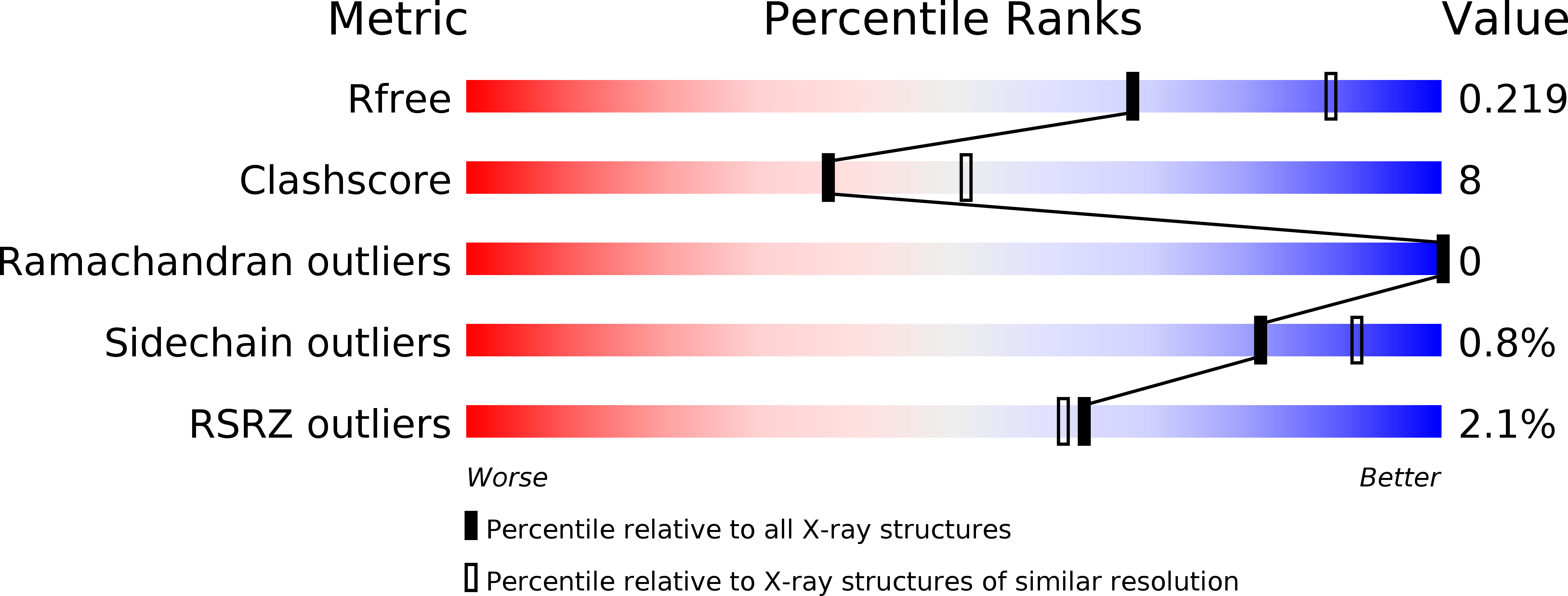
Resolution:
2.40 Å
R-Value Free:
0.22
R-Value Work:
0.17
R-Value Observed:
0.18
Space Group:
P 21 21 2
Macromolecular Entities
Structures with similar UniProt ID
Protein Blast
Polymer Type:polypeptide(L)
Molecule:DELTA TOXIN
Chain IDs:A
Chain Length:298
Number of Molecules:1
Biological Source:CLOSTRIDIUM PERFRINGENS
Primary Citation
Structural Insights Into Clostridium Perfringens Delta Toxin Pore Formation.
Plos One 8 66673 ? (2013)
PMID: 23805259 DOI: 10.1371/JOURNAL.PONE.0066673

Abstact

Clostridium perfringens Delta toxin is one of the three hemolysin-like proteins produced by C. perfringens type C and possibly type B strains. One of the others, NetB, has been shown to be the major cause of Avian Nectrotic Enteritis, which following the reduction in use of antibiotics as growth promoters, has become an emerging disease of industrial poultry. Delta toxin itself is cytotoxic to the wide range of human and animal macrophages and platelets that present GM2 ganglioside on their membranes. It has sequence similarity with Staphylococcus aureus β-pore forming toxins and is expected to heptamerize and form pores in the lipid bilayer of host cell membranes. Nevertheless, its exact mode of action remains undetermined. Here we report the 2.4 Å crystal structure of monomeric Delta toxin. The superposition of this structure with the structure of the phospholipid-bound F component of S. aureus leucocidin (LukF) revealed that the glycerol molecules bound to Delta toxin and the phospholipids in LukF are accommodated in the same hydrophobic clefts, corresponding to where the toxin is expected to latch onto the membrane, though the binding sites show significant differences. From structure-based sequence alignment with the known structure of staphylococcal α-hemolysin, a model of the Delta toxin pore form has been built. Using electron microscopy, we have validated our model and characterized the Delta toxin pore on liposomes. These results highlight both similarities and differences in the mechanism of Delta toxin (and by extension NetB) cytotoxicity from that of the staphylococcal pore-forming toxins.

Legend

Protein

Chemical

Disease

Primary Citation of related structures
Feedback Form
Name
Email
Institute
Feedback