Deposition Date
2011-04-18
Release Date
2011-06-22
Last Version Date
2023-12-20
Entry Detail
PDB ID:
2YGL
Keywords:
Title:
The X-ray crystal structure of tandem CBM51 modules of Sp3GH98, the family 98 glycoside hydrolase from Streptococcus pneumoniae SP3-BS71
Biological Source:
Source Organism(s):
STREPTOCOCCUS PNEUMONIAE (Taxon ID: 406556)
Expression System(s):
Method Details:
Experimental Method:
Resolution:
2.10 Å
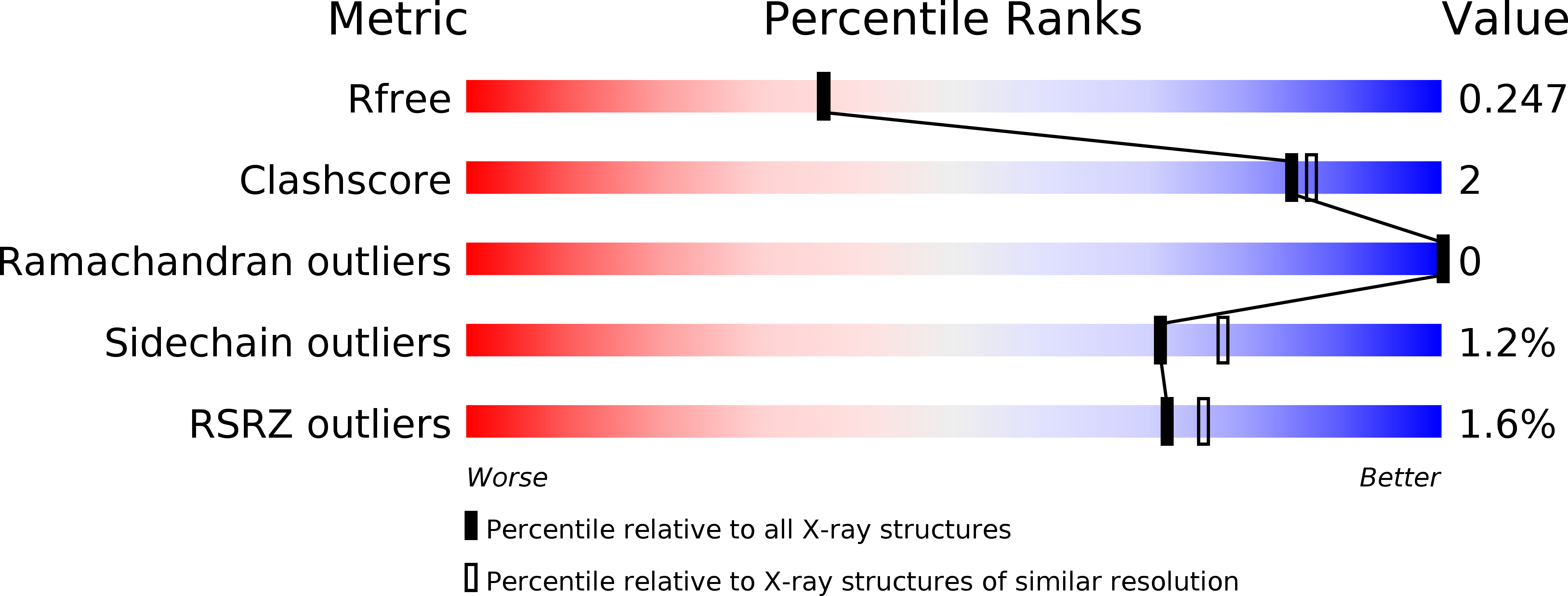
R-Value Free:
0.24
R-Value Work:
0.20
R-Value Observed:
0.21
Space Group:
P 21 21 21