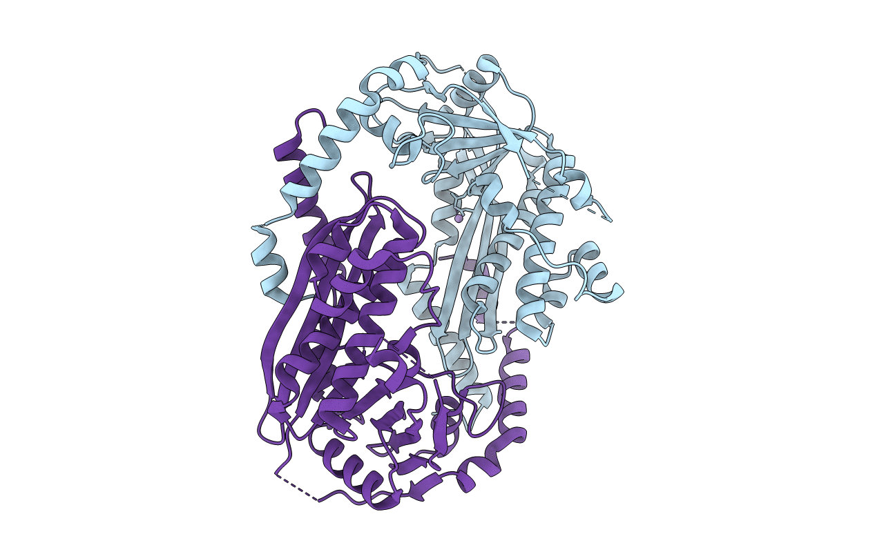
2YGK image
Deposition Date 2011-04-18
Release Date 2011-12-14
Last Version Date 2024-10-09
Entry Detail
PDB ID:
2YGK
Keywords:
Title:
Crystal structure of the NurA nuclease from Sulfolobus solfataricus
Biological Source:
Source Organism:
Host Organism:
Method Details:
Experimental Method:
Resolution:
2.50 Å
R-Value Free:
0.27
R-Value Work:
0.23
R-Value Observed:
0.23
Space Group:
P 1 21 1
Macromolecular Entities
Polymer Type:polypeptide(L)
Molecule:NURA
Gene (Uniprot):nurA
Chain IDs:A, B
Chain Length:339
Number of Molecules:2
Biological Source:SULFOLOBUS SOLFATARICUS
Modified Residue
Compound ID Chain ID Parent Comp ID Details 2D Image
MSE A MET SELENOMETHIONINE
Ligand Molecules
Primary Citation
Structural and Functional Insights Into DNA-End Processing by the Archaeal Hera Helicase-Nura Nuclease Complex.
Nucleic Acids Res. 40 3183 ? (2012)
PMID: 22135300 DOI: 10.1093/NAR/GKR1157

Abstact

Helicase-nuclease systems dedicated to DNA end resection in preparation for homologous recombination (HR) are present in all kingdoms of life. In thermophilic archaea, the HerA helicase and NurA nuclease cooperate with the highly conserved Mre11 and Rad50 proteins during HR-dependent DNA repair. Here we show that HerA and NurA must interact in a complex with specific subunit stoichiometry to process DNA ends efficiently. We determine crystallographically that NurA folds in a toroidal dimer of intertwined RNaseH-like domains. The central channel of the NurA dimer is too narrow for double-stranded DNA but appears well suited to accommodate one or two strands of an unwound duplex. We map a critical interface of the complex to an exposed hydrophobic epitope of NurA abutting the active site. Based upon the presented evidence, we propose alternative mechanisms of DNA end processing by the HerA-NurA complex.

Legend

Protein

Chemical

Disease

Primary Citation of related structures