Deposition Date
2011-04-15
Release Date
2011-09-28
Last Version Date
2024-05-08
Entry Detail
Biological Source:
Source Organism(s):
HOMO SAPIENS (Taxon ID: 9606)
RATTUS NORVEGICUS (Taxon ID: 10116)
RATTUS NORVEGICUS (Taxon ID: 10116)
Expression System(s):
Method Details:
Experimental Method:
Resolution:
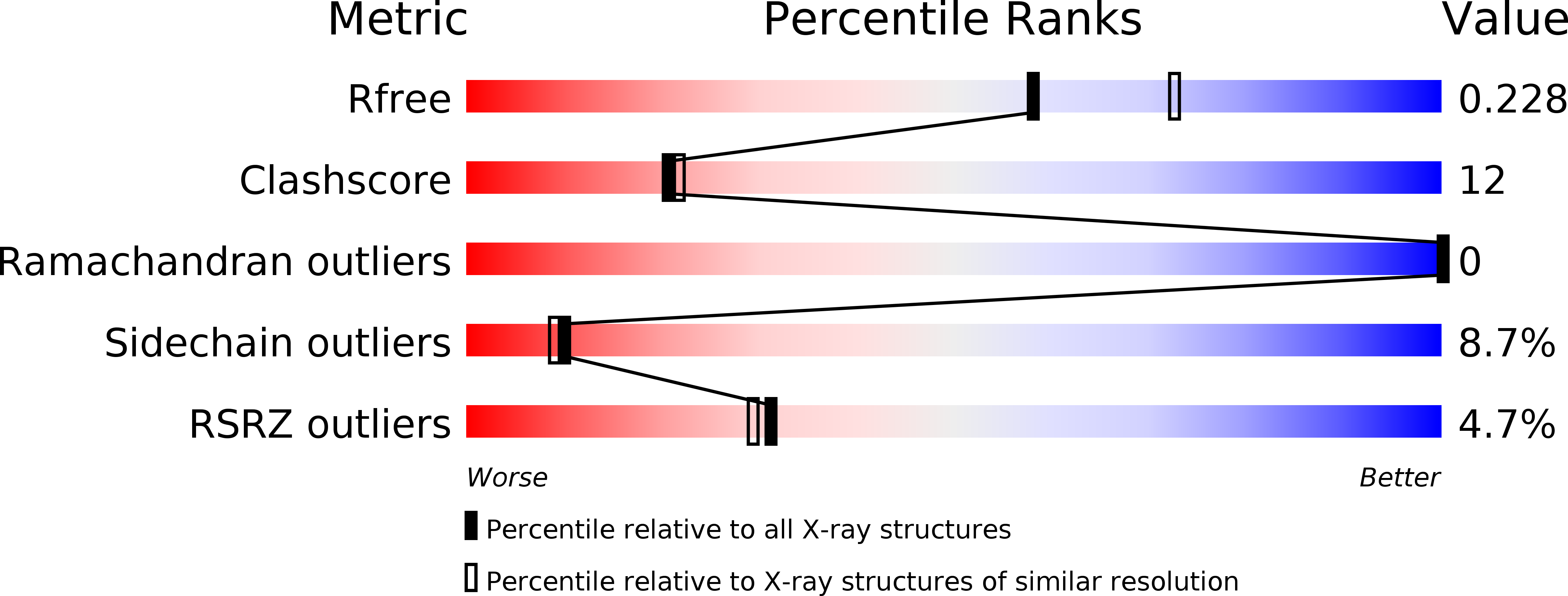
2.23 Å
R-Value Free:
0.23
R-Value Work:
0.17
R-Value Observed:
0.17
Space Group:
C 1 2 1